Enumerate the layers of anterior abdominal wall.
Layers of anterior abdominal wall are:
- Skin
- Superficial fascia
- Outer fatty layer (Camper’s fascia)
- Inner membranous layer (Scarpa’s fascia)
- Muscles (arranged in three layers)
- External oblique
- Internal oblique
- Transversus abdominis
- Fascia transversalis
- Extraperitoneal tissue
- Peritoneum (parietal layer)
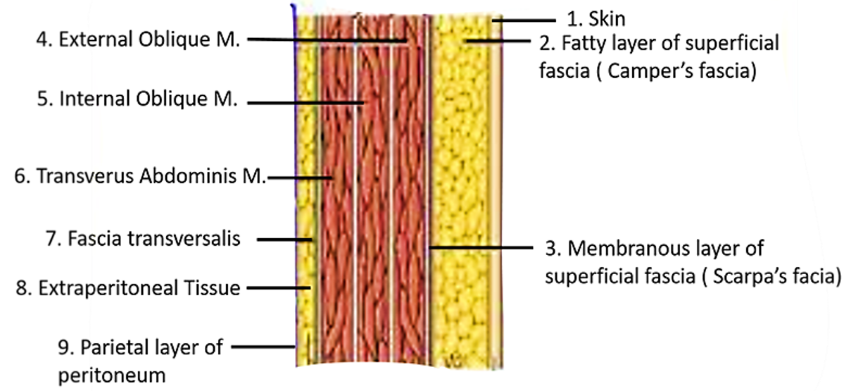
There is no deep fascia in the anterior abdominal wall.
Name the planes used for dividing abdominal cavity into regions.
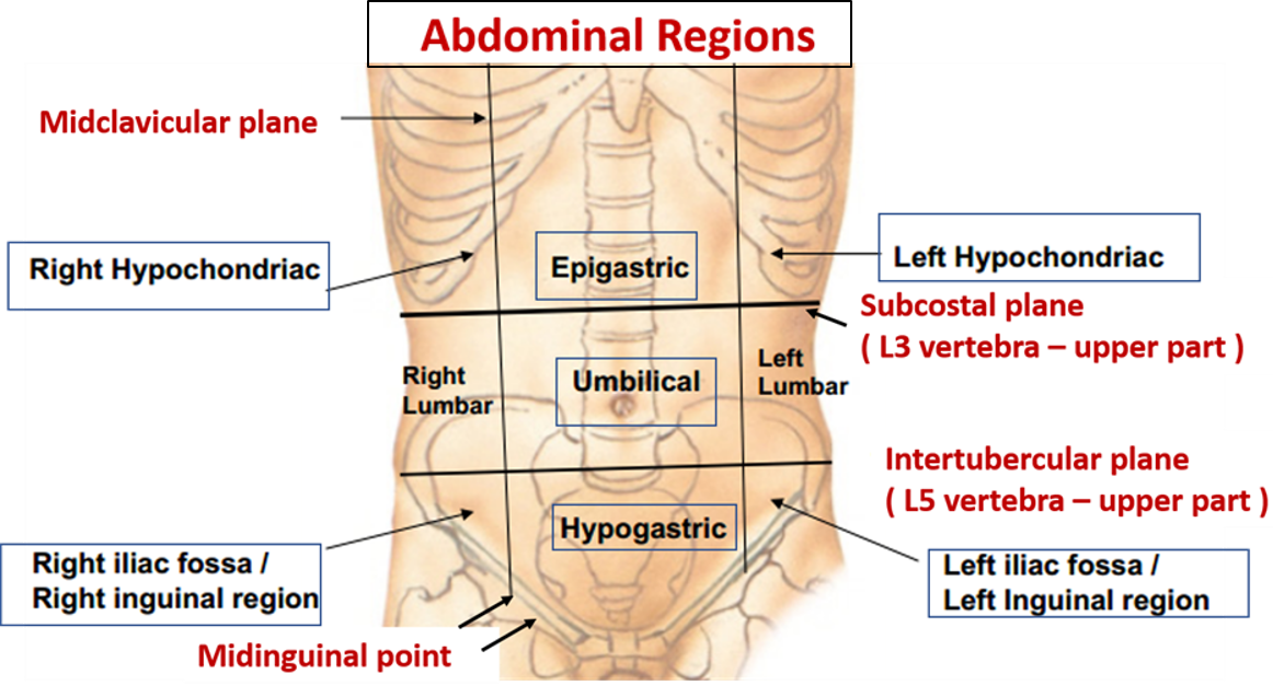
Abdomen is divided into nine regions using:
- Two horizontal planes:
- Transpyloric plane: passes in front through the tips of the ninth costal cartilages and behind at the lower border of L1 vertebra.
- Subcostal plane: passes in front at the level of inferior margin of 10th rib and behind through the upper part of L3 vertebra.
- Two vertical planes:
- Right and left midclavicular planes: pass through the midpoint of clavicle to the midinguinal point (midway between the anterior superior iliac spine and pubic symphysis.
What are the vertebral levels of important abdominal planes?
- Transpyloric plane: Lower border of L1 vertebra
- Subcostal plane: Upper part of body of L3 vertebra
- Plane passing through the highest point of iliac crest: L4 Vertebra
- Intertubercular plane – Upper part of body of L5 vertebra
Enumerate the structures present at the transpyloric plane.
- Pylorus of stomach
- Neck of pancreas
- Duodeno – jejunal flexure
- Fundus of gall bladder
- Hilum of kidneys
- Origin of superior mesenteric artery
- Tip of ninth costal cartilage
- Termination of spinal cord
Name the nine abdominal regions and their main contents.
Region Contents Region Contents Region Contents Right hypochondrium Right lobe of liver Epigastrium Pyloric part of stomach Left hypochondrium Spleen Gall bladder Liver Tail of pancreas Right colic flexure Pancreas Stomach Right suprarenal gland Duodenum Left suprarenal gland Upper part of right kidney Abdominal aorta Upper part of left kidney Left colic flexure Right lumbar region Lower part of right kidney & ureter Umbilical region Abdominal aorta Left lumbar region Lower part of the kidney, ureter Ascending colon Inferior vena-cava Descending colon Part of duodenum Stomach and greater omentum Coils of jejunum Coils of jejunum Transverse colon Jejunum & ileum Right iliac region Terminal part of ileum Hypogastrium Urinary bladder (when distended), Left iliac region Sigmoid colon Caecum with appendix Coils of ileum, Left ureter Ascending colon Gravid uterus in pregnant mother Right ureter
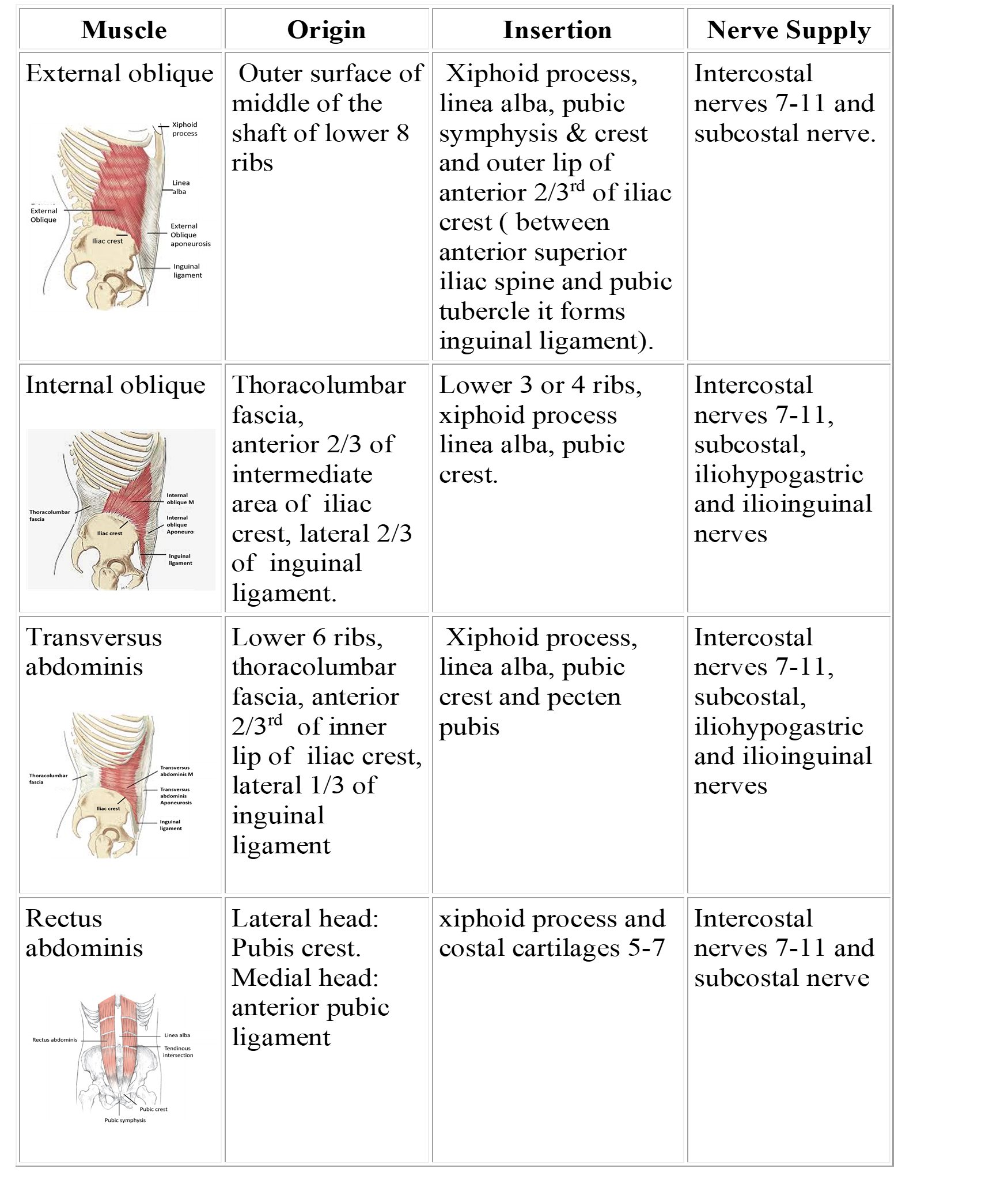
Write the origin, insertion and nerve supply of muscles of anterior abdominal wall.
What are the action/s of anterior abdominal wall muscles?
Anterior abdominal wall muscles
- Support and protect the abdominal viscera.
- Help in forced expiration that occurs during coughing, sneezing, vomiting,
- Increase the intra-abdominal pressure and thereby help in defecation, micturition (urination), and parturition (childbirth).
- Rotate the trunk to the opposite side (external oblique of one side and internal oblique of the opposite side).
- Flex the trunk laterally (external and internal oblique muscles of the same side).
- Flex the trunk anteriorly (rectus abdominis).
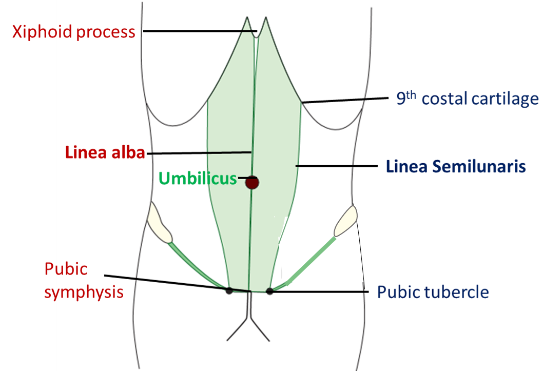
What are linea alba, linea semilunaris?
Linea alba
- Is an avascular fibro-tendinous raphe formed by the interlacing fibers of the aponeurosis of the three lateral muscles of anterior abdominal wall (external oblique, internal oblique and transversus abdominis).
- It extends from the xiphoid process to pubic symphysis.
Linea semilunaris
- A curved fibrous line at the lateral margin of the rectus sheath that extends from the tip of the ninth costal cartilage to the pubic tubercle.
Write a short note on Umbilicus.
- Is a depressed scar in the midline of anterior abdominal wall , normally between the xhiphoid process and pubic symphysis or between L3 and L4 vertebra.
- Represents the site of attachment of fetal end of umbilical cord.
- Is innervated by T10 spinal segment.
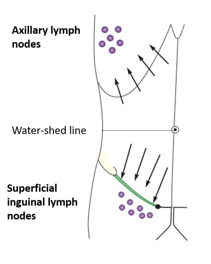
- Acts as a water – shed line with respect to lymph and venous flow.
- Is one of the sites of portocaval anastomosis.
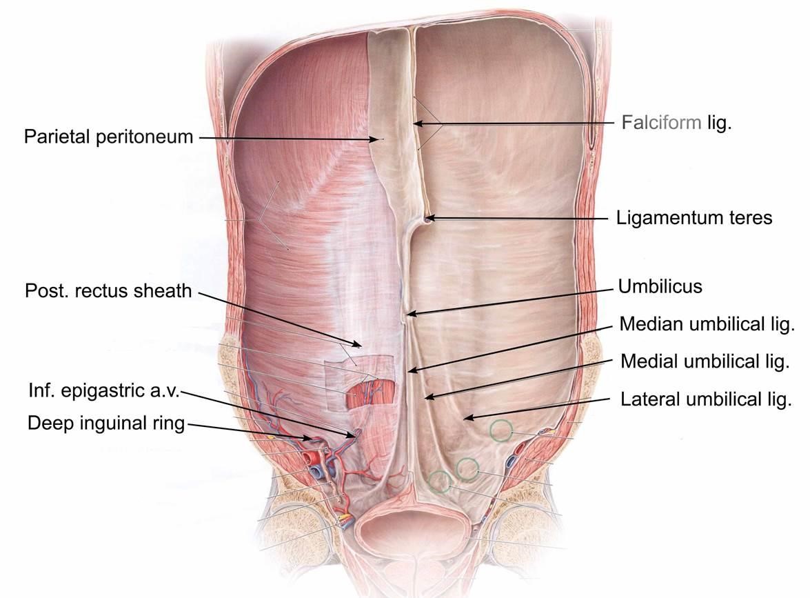
- Fibrous bands attached to the inner aspect of umbilicus:
- Ligamentum teres hepatis – remnant of left umbilical vein
- Median umbilical ligament (Urachus)– remnant of allantois
- Medial umbilical ligaments – remnant of umbilical arteries
Applied Aspect
Omphalocele
Occurs due to failure of reduction of physiological herniation of midgut loop, herniated loops of small intestine are covered by amnion.
Congenital umbilical hernia
Occurs due to herniation of loops of small intestine through imperfectly closed umbilicus. The herniated loop is covered by peritoneum.
Fecal fistula
Occurs due to persistence and complete patency of vitellointestinal duct through which ileum communicates with the exterior.
Urinary fistula
Occurs due to patent urachus (connecting umbilicus to urinary bladder).It may lead to dribbling of urine from the umbilicus.
Caput medusae
In case of portal hypertension, the superficial veins radiating from the umbilicus (site of portocaval anastomosis) become dilated and tortuous.
Referred pain in appendicitis
Referred pain in case of appendicitis is felt at umbilicus as both are supplied by T10 spinal segment.
Describe the superficial fascia of anterior abdominal wall.
Superficial fascia of anterior abdominal wall: In the lower part of the anterior abdominal wall (below the line passing through the midpoint between the umbilicus and pubic symphysis), the superficial fascia has two layers:
- Superficial fatty layer (Camper’s fascia)
- Deep membranous layer (Scarpa’s fascia)
The superficial fatty layer is continuous with the superficial fascia of the rest of the body, the membranous layer is devoid of fat and has more of elastic fibers.
Attachment of Scarpa’s fascia:
- From the lower half of anterior abdominal wall, it passes over the inguinal ligament and is attached to the deep fascia of the thigh along an horizontal line (Holden’s line approx. 8cm.) passing laterally from the pubic tubercle.
- Medially it passes downwards over the body of the pubis and becomes continuous with the superficial perineal fascia (Colle’s fascia). The superficial perineal fascia is attached laterally to conjoint ischiopubic rami and posteriorly it is fused with the posterior border of perineal membrane.
- It also extends into the penis and scrotum (labia majora in females).
Applied Aspect
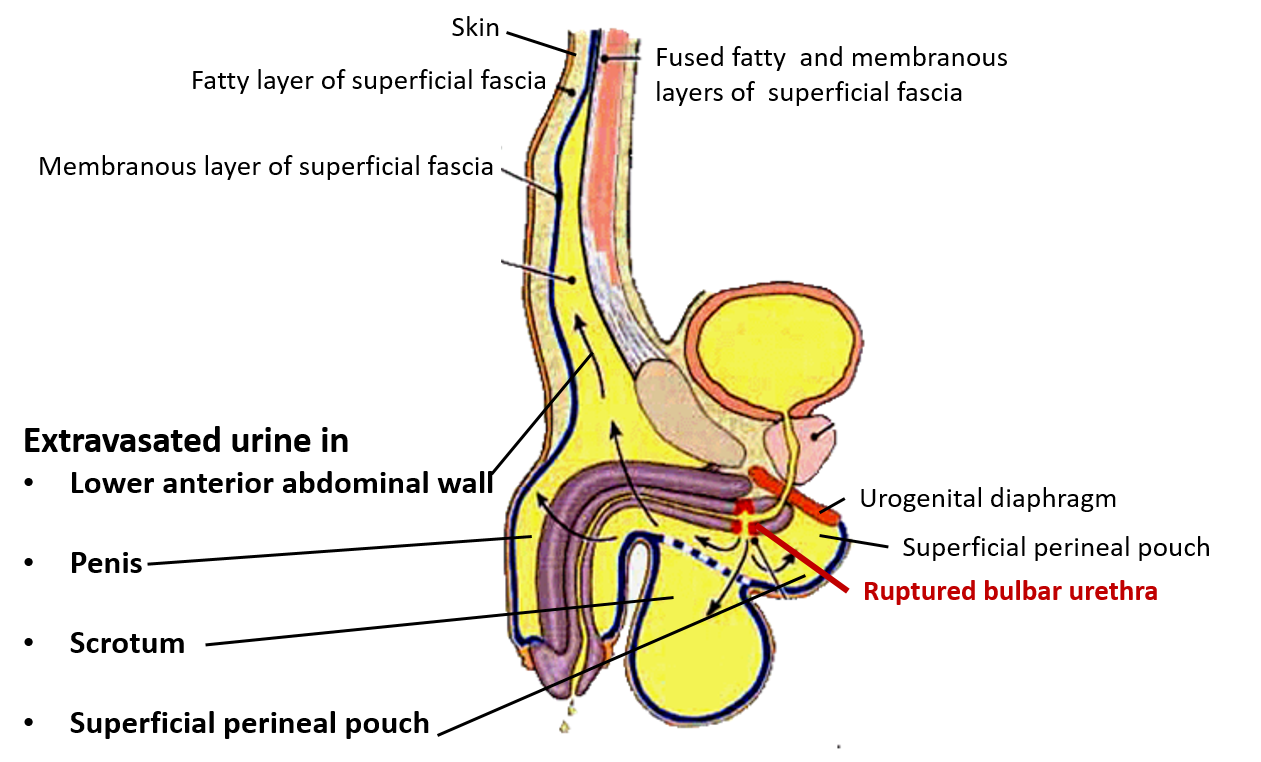
Course of extravasated urine in case of rupture of bulbar urethra
If bulbar urethra is ruptured, the extravasated urine first fills the superficial perineal pouch of perineum and then extends to the scrotum, penis and the anterior abdominal wall (inferior to the level of umbilicus), deep to the Scarpa’s fascia. But, the urine will not extend into the thigh due to the attachment of membranous layer of superficial fascia to fascia lata of thigh along the Holden’s line, body of the pubis and pubic arch.
Enumerate the arteries that supply anterior abdominal wall.
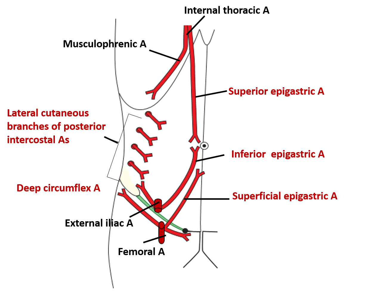
The following arteries supply anterior abdominal wall:
- Superior epigastric, branch of internal thoracic artery.
- Lateral cutaneous branches of posterior intercostal arteries.
- Superficial epigastric , branch of femoral artery
- Inferior epigastric – branch of external iliac artery.
- Deep circumfles, branch of external iliac artery.
Enumerate the cutaneous nerves innervating anterior abdominal wall.
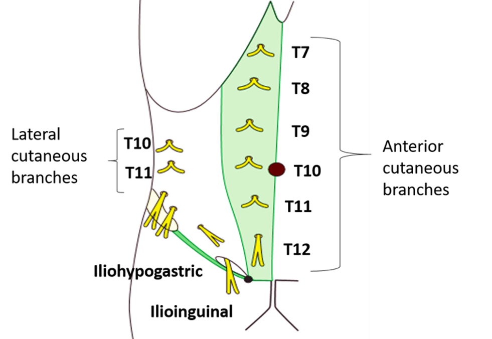
Following cutaneous nerves supply anterior abdominal wall:
- Anterior cutaneous branches of T7-T11 intercostal nerves and subcosatl nerve.
- Lateral cutaneous branches of T10 and T11 intercostal nerves.
- Iliohypogastric nerve.
Describe in brief the lymphatic drainage of anterior abdominal wall.
- Lymphatics from anterior abdominal wall above the level of umbilicus drains into axillary lymph nodes.
- Lymphatics from anterior abdominal wall below the level of umbilicus drains into superficial inguinal lymph nodes.

two horizontal planes dividing the abdomen into 9 regions is done mistake
The Anatomy described and explained very good thanks you.
I have benefited a lot here