What are the Parts of Ear?
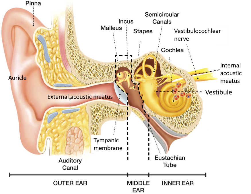
The ear consists of following three parts:
External ear
It consists of auricle (pinna) and external acoustic meatus(EAM). The auricle collects the sound waves and EAM conducts them to the tympanic membrane. External ear is separated from the middle ear by tympanic membrane.
Middle ear
It is an air-filled cavity which is separated from internal ear by a bony wall. It communicates anteriorly with the nasopharynx through the auditory tube (Eustachian tube).
Internal ear
Internal ear contains bony labyrinth (cochlea, vestibule and semicircular canals) within which lies membranous labyrinth containing receptors for sound and maintenance of equilibrium. Vetsibulocochlear nerve reaches internal ear through the internal acoustic meatus.

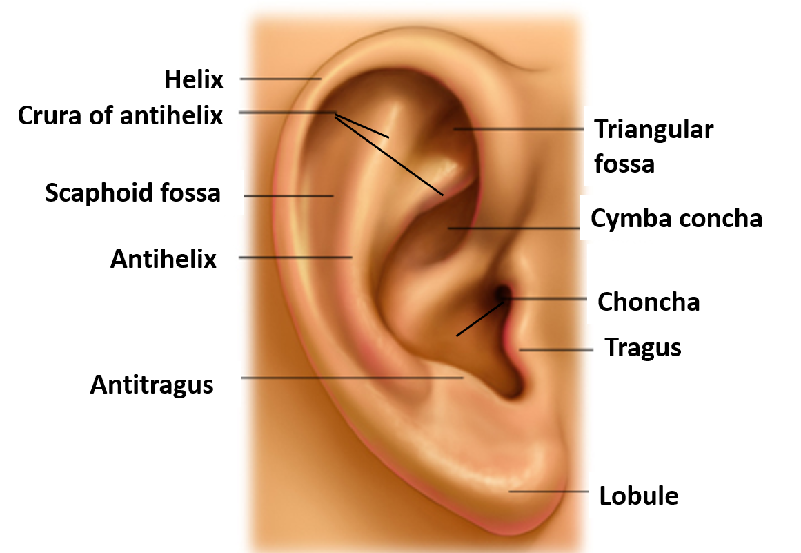
Draw a Labelled Diagram to Show Gross Features of Auricle.

Name the Sensory nerves Supplying Auricle.
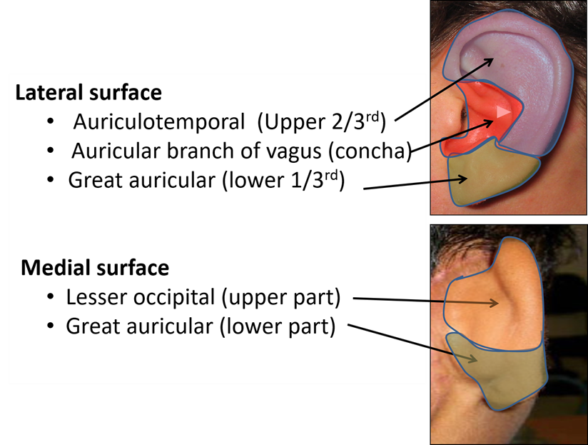
Sensory innervation of auricle: The following nerves supply the skin of auricle:
- Great auricular nerve (ventral rami of C2,C3): supplies lower 1/3 of the auricle/pinna on lateral and medial surfaces of auricle.
- Auriculotemporal nerve (branch of posterior division of mandibular nerve): supplies upper 2/3rd of the lateral surface of auricle.
- Lesser occipital (ventral ramus of C2): supplies upper 2/3rd of the medial/cranial surface of auricle.
- Auricular branch of vagus: supplies the root of auricle (near external acoustic meatus).

What are the Parts of External Acoustic Meatus?
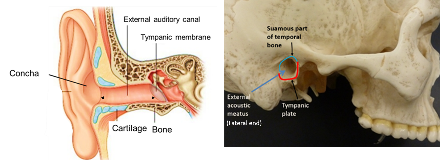
The external acoustic/auditory meatus (EAM):
- Extends from concha to tympanic membrane.
- It is approx. 24-25 mm long.
- It has two parts-
- Outer cartilaginous part: it is 8 mm long. Its wall contains elastic cartilage.
- Inner bony part: it is 16 mm long. Tympanic plate of temporal bone forms its anterior, inferior and posterior walls. Posterosuperior wall is formed by squamous part of temporal bone.
- At the medial end of the bony canal is tympanic membrane.
- EAM has two constrictions:
- First is at the junction of cartilaginous and bony part.
- Second called isthmus is its narrowest part, which is located 6 mm lateral to tympanic membrane.
- It is lined with skin and has numerous ceruminous gland which secret ear wax.

Shape and Direction: It is S-shaped and has two curves.
- Outer part is directed upwards, forwards and medially.
- Middle part is directed upwards, backwards and medially.
- Inner part is directed downwards, forwards and medially
Name the Sensory Nerves That Supply External Acoustic Meatus.
Sensory innervation of EAM is by following nerves:
- Auriculotemporal nerve– supplies roof and anterior wall.
- Auricular branch of vagus– supplies floor and posterior wall.
Applied Aspects
During examination of ear, the auricle is pulled upwards, backwards, outwards
As external auditory meatus is ‘S’ shaped, therefore, during examination of ear, the auricle is pulled upwards, backwards, outwards in adults, to straighten the ear canal to a certain extent and provide a better view in examination.
Syringing of external auditory meatus to remove wax may stimulate auricular branch of vagus
Skin over the concha of external ear is supplied by auricular branch of vagus and therefore, syringing of external auditory meatus to remove wax may stimulate auricular branch of vagus and result in:
- reflex coughing
- vomiting
- may result in cardiac arrest also.
