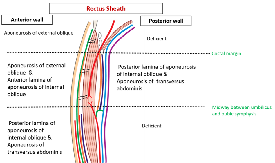
Describe the formation of rectus sheath.
Rectus sheath
- is an aponeurotic envelope for rectus abdominis and pyramidalis muscles.
- is situated on either side of linea alba.
- strengthens the anterior abdominal wall and prevents the bow stringing of the rectus abdominis muscle.
- has anterior and posterior walls and medial and lateral margins.
Formation:
- Anterior wall: It is complete but differs in thickness at different levels.
- In the region above the costal margin: It is thin and formed by aponeurosis of external oblique.
- In the region from costal margin to midway between umbilicus and pubic symphysis: Is thicker and is formed by the anterior lamella of aponeurosis of internal oblique with the aponeurosis of external oblique.
- In the region between the midpoint of umbilicus and pubic symphysis to pubic symphysis: Is thickest and is formed by the fusion of the aponeurosis of the three flat muscles of the abdomen i.e external oblique, internal oblique and transversus abdominis.
* Anterior wall of the rectus sheath is adherent to the anterior surface of the rectus muscle along the three tendinous intersections.
- Posterior wall:
- In the region above the costal margin: In this region the posterior wall is deficient and the rectus abdominis rests directly on the 5th, 6th and 7th costal cartilages. The posterior wall of the rectus sheath is deficient here because the transversus abdominis muscle passes internal to the costal cartilages and the internal oblique muscle is attached to the costal margin.
- In the region from the costal margin to midway between umbilicus and symphysis pubis: It is formed by the posterior lamella of aponeurosis of internal oblique with the aponeurosis of transversus\ abdominis. A crescentic border called the arcuate line marks the inferior limit of the posterior wall of the rectus sheath. It is usually midway between the umbilicus and the pubic crest.
- In this region between the midpoint of umbilicus and pubic symphysis to pubic symphysis : It is formed by fascia transversalis. Inferior to the arcuate line, the aponeurosis of the three flat muscles pass anterior to the rectus muscle to form the anterior layer of the rectus sheath.


Sagittal Section Through Rectus Sheath

Enumerate the contents of rectus sheath.
Muscles
- Rectus abdominis
- Pyramidalis
Blood vessels
- Superior epigastric
- Inferior epigastric
Nerves
- Terminal parts of lower 5 intercostals
- Subcostal
Applied Aspects
Anatomical Basis of Abdominal Incisions
Midline incision
It is made through linea alba. Linea alba provides an almost bloodless line along which abdomen can be opened. The disadvantage is that it leads to post operative weakness and herniation may occur.
Paramedian incision: Is placed 1-1 ½ inch lateral and parallel to the midline, the anterior wall of rectus sheath is opened, the rectus abdominis is displaced laterally (to avoid injury to the nerves supplying rectus abdominis) and the posterior wall of rectus sheath along with fascia tranversalis and the peritoneum is then incised.
*Rectus abdominis should be retracted laterally to avoid injury to nerves that pierce the sheath laterally.
Subcostal (Kocher) incision
It is used on the right side forgall bladder surgery and on the left, for exposure of spleen. The skin incision begins at the midline and extends parallel to and 1 inch below the costal margin.
Muscle split or grid iron incision (McBburney’s incision)
A transverse incision in theline of the skin crease 1 inch above and forward from the anterior superior iliac spine or an oblique incision centered at Mc Burney’s point along the line from anterior superior iliac spine to umbilicus. The aponeurosis of external oblique is incised obliquely downwards and medially (in the line of its fibers). The internal oblique and the transversus muscles are then split in the line of their fibers and retracted without dividing their fibers. On closing the incision, these muscles snap together, leaving almost undamaged anterior abdominal wall.


Thanks a lot……your work is very organized