Enumerate Contents of the Orbit.
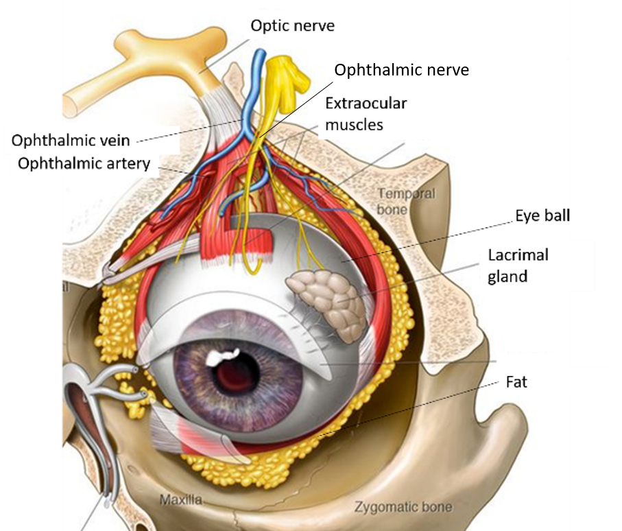
Contents of orbit are:
- Eyeball
- Extraocular muscles
- Nerves: oculomotor, trochlear, abducent, and three branches of ophthalmic nerve i.e. frontal, nasocilliary and lacrimal nerves.
- Blood vessels: ophthalmic artery and its branches, superior and inferior ophthalmic veins.
- Lacrimal gland
- Fat

Write the Origin and Insertion of Extraocular Muscles.
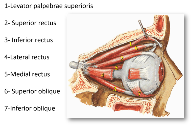
The muscle acting on the eye ball to produce various movements of eye are called extraocular muscles. These are:
- Four recti muscles: superior rectus, inferior rectus, medial rectus & lateral rectus.
- Two oblique muscles: superior and inferior oblique muscles.
- Levator palpabrae superioris: This muscle does not act on eyeball, but is responsible for elevating the upper eyelid. Following are the extraocular muscles:

Recti Muscles
- Origin: All the four recti muscles take origin from the corresponding margins of a common fibrous ring (tendon of Zinn) which encircles optic canal and adjacent part of superior orbital fissure.
- Insertion: All recti are inserted into sclera in front of the equator, about 5.5-7mm behind the sclerocorneal junction/limbus.

Oblique muscles
Superior oblique:
- Origin – from body of sphenoid, just above the optic canal. The muscle passes along superomedial aspect of orbit and then turns in posterolateral direction by passing through a fibrocartilaginous ring (attached at the anteromedial part of roof of orbit).
- Insertion – Into sclera in the superolateral quadrant of eyeball.
Inferior oblique:
- Origin – Into sclera in the anteromedial part of floor of orbit.
- Insertion- In to sclera in the infero-lateral quadrant of eyeball.
Levator palpebral superioris
- Origin: It arises from lesser wing of sphenoid just above the common tendinous ring.
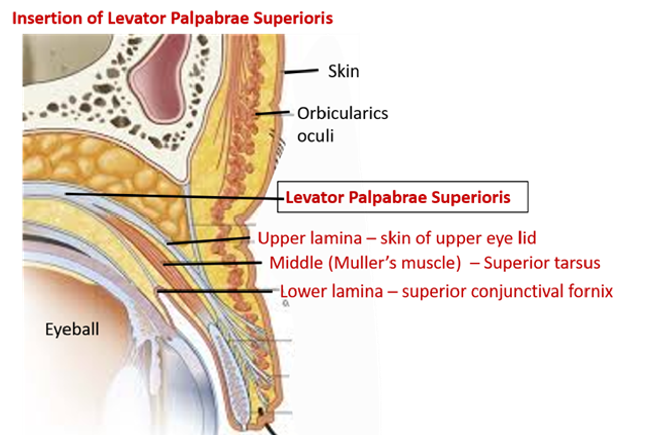
- Insertion: Aponeurosis of the muscle splits into three lamina:
- Upper lamina is inserted into skin of upper eyelid.
- Middle lamina containing smooth muscle (Muller’s muscle) is attached to upper margin of superior tarsus.
- Lower lamina is attached to superior conjunctival fornix.

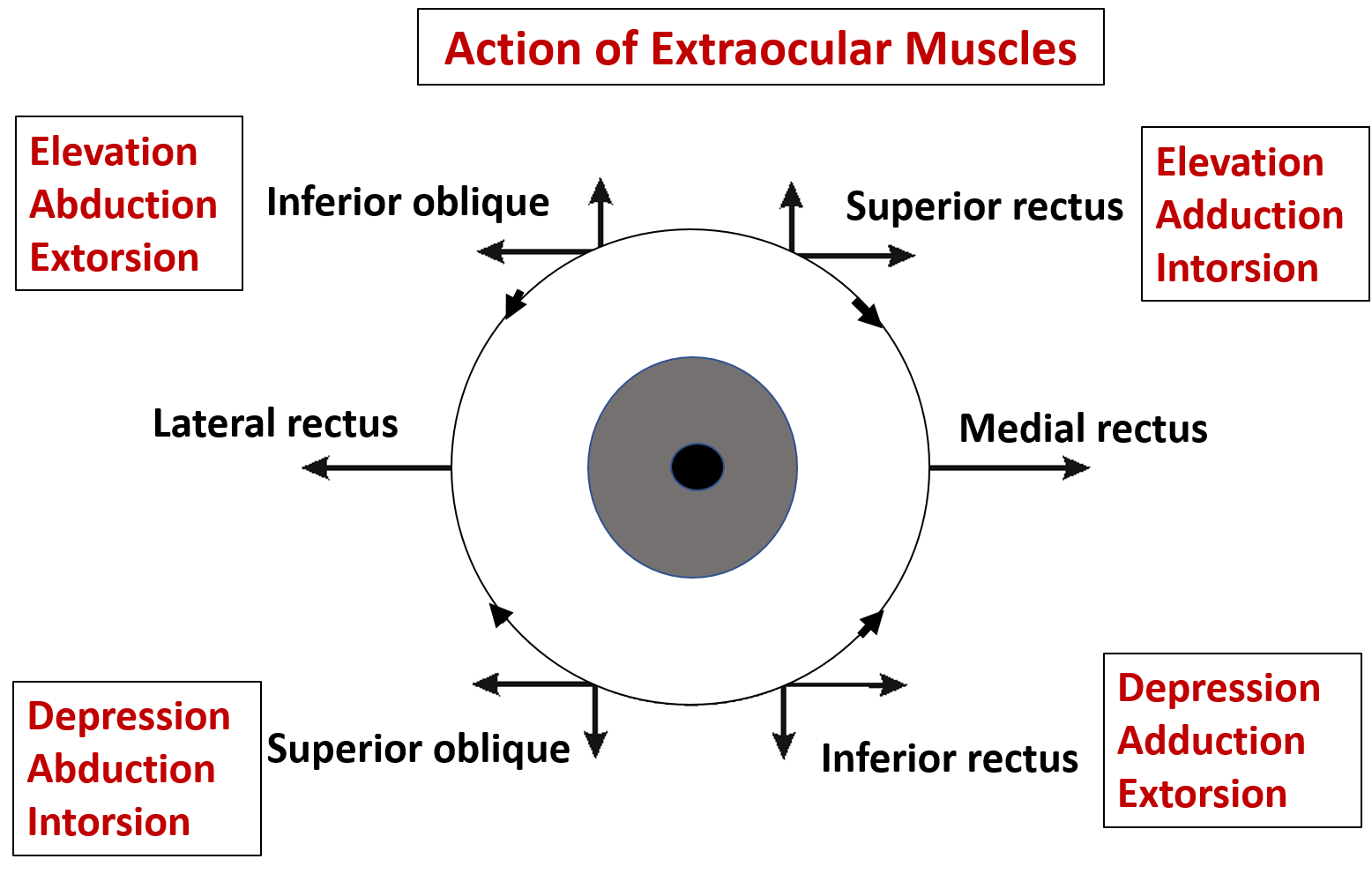
What is the action and nerve supply of Extraocular Muscles?
| Muscle | Action | Nerve supply |
|---|---|---|
| Superior rectus | Elevation ,adduction and intorsion | Oculomotor N |
| Inferior rectus | Depression adduction and extorsion | Oculomotor N |
| Lateral rectus | Abduction | Abducent N |
| Medial rectus | Adduction | Oculomotor N |
| Superior oblique | Depression, abduction & intorsion | Trochlear N |
| Inferior oblique | Elevation , abduction & extorsion | Oculomotor N |
| Levator palpabrae superioris | Elevation of upper eyelid | Oculomotor and sympathetic fibers from T1 spinal segment. |

Enumerate the Structures Passing Through the Superior Orbital Fissure
Superior orbital fissure (SOF) is divided into three parts i.e . upper, middle and lower parts by the Annulus of Zinn/tendinous ring. The structures passing through the three parts of SOF are:
Through the Upper part:
- lacrimal nerve
- frontal nerve
- trochlear nerve
- superior ophthalmic vein
Through the Middle part:
- superior and inferior division of oculomotor nerve
- abducens nerve,
- nasociliary nerve
Through the Lower part:
- inferior ophthalmic vein

For
Ophthalmic nerve [Click here]
Ophthalmic artery [Click here]
Ciliary ganglion [Click here]
Applied Aspects
Strabismus/squint
Unilateral paralysis of lateral or medial rectus muscle results in deviation of eye to the opposite side.
- Internal or medial squint: paralysis of lateral rectus muscle.
- External or lateral squint: paralysis of medial rectus muscle.
Paralysis of superior oblique muscle
It causes diplopia on looking down as in walking down the stairs.
Ptosis
It means drooping of upper eyelid. It can occur due to:
- Paralysis of levator palpebrae superioris due to injury to oculomotor nerve.
- Paralysis of Muller’s muscle (part of levator palpabrae superioris supplied by sympathetic fibers) due to injury to sympathetic fibers supplying it (as in Horner’s syndrome).
