Introduction
Mammary gland is a modified sweat gland situated in the superficial fascia of pectoral region. A small extension called axillary tail of Spence pierces deep fascia and lies in the axilla.
- Extent: Vertically from 2nd to 6th rib and horizontally from lateral border of the sternum to the mid-axillary line.
- Deep relations: From superficial to deep are:
- Loose areolar tissue (retromammary space)
- Deep fascia (pectoral fascia)
- Three muscles – pectoralis major, serratus anterior and external oblique
Describe the Structure of Mammary gland.
Mammary gland is made up of the following two components:
- Parenchyma (glandular tissue): It is made up of glandular tissue comprising 15 to 20 radially arranged in pyramidal lobes. Each lobe has a cluster of alveoli which is drained by a lactiferous duct. Lactiferous ducts open on the nipple and just before its termination each duct is dilated to form lactiferous sinus.
- Stroma (fibrofatty tissue): Fatty tissue forms the main bulk of the gland but is absent beneath the areola and nipple. The fibrous septa (suspensory or Cooper’s ligament) anchor skin overlying the gland and the gland to the pectoral fascia.

Name the arteries Suppling mammary Gland
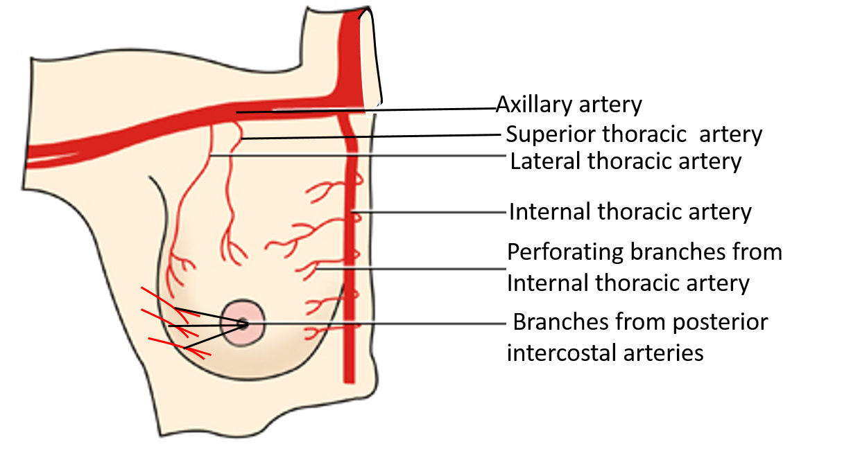
Following arteries supply the mammary gland:
- Perforating branches of internal thoracic artery.
- Branches of axillary artery (superior thoracic, thoracoacromial and lateral thoracic).
- Lateral branches of 2nd, 3rd and 4th posterior intercostals arteries.

Describe the Lymphatic Drainage of Mammary Gland.
Lymphatic drainage of mammary gland is of great clinical significance because carcinoma of breast spreads mainly along the lymphatics.Lymphatic vessels of the breast are arranged into two groups:
- Superficial lymphatic vessels drain the lymph from the overlying skin except nipple and areola.
- Deep lymphatic vessels drain the parenchyma along with nipple and areola.
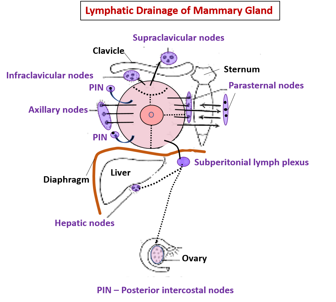
Lymph from the mammary gland is drained into the following groups of lymph nodes:
i. 75% of the lymph drains into axillary lymph nodes.
ii. 20% of the lymph drains into internal mammary (parasternal) lymph nodes.
iii. 5% of the lymph drains into posterior intercostal lymph nodes.
iv. Lymph from superior quadrants drains into supraclavicular lymph nodes.
v. Lymph vessels from infero-medial quadrant communicate with the subperitoneal lymph plexus.

Applied Aspects
Peau d’orange appearance
Obstruction of superficial lymph vessels leads to stagnation of lymph resulting in odema of skin (peau d’orange appearance – like the skin of orange).
Rretraction or puckering of skin over mammary gland.
Cancer cells may infiltrate the suspensory ligaments resulting in fixation of breast to pectoral fascia and retraction or puckering of skin.
Retraction of nipple.
Infiltration of lactiferous ducts by cancer cells leads to fibrosis of lactiferous ducts which causes retraction of nipple.
Cancer may spread from one breast to the other.
Because of the communication between the superficial lymphatics across the midline.
Krukenberg’s tumour
Lymph vessels from inferomedial quadrant communicate with with subperitoneal lymph plexus.Cancer cells therefore may spread to the liver may drop into the pelvis and produce secondary tumour in the ovary ( Krukenberg’s tumour).
Cancer from breast may spread the brain.
Besides lymphatics cancer may spread via the veins. Intercostal veins draining mammary gland communicate with the internal vertebral venous plexus which in turn communicates with the basilar plexus of veins in the cranial cavity. Therefore, cancer from breast may spread via this communication to the vertebrae and to the brain.
Breast abcess is drained by radial incisions.
Beacuse the lactiferous ducts are arranged radially around the nipple and therefore, to avoiding cutting across lactiferous ducts.

Thanks
Pretty good 🙂🙂😊😊
But miss information about the areola
Pretty good
I do really love this very much. Please O want it in subcopy if possible