Advertisements
What are the components of conducting system of heart?
Conducting system of heart is meant for initiating and maintaining cardiac rhythm and establish proper co-ordination between the atrial and ventricular contactions. It is made up of specialized cardiac muscle fibers having a high degree of sensitivity and autorhythmicity. Following are the components of conducting system: 
1. Sinuatrial node (SA node)
- Is also known as pacemaker.
- Initiates the cardiac impulse.
- Is located in the upper part of crista terminalis by the side of the opening of superior vena cava.
- Impulse from SA node to AV node is carried by internodal fibers.
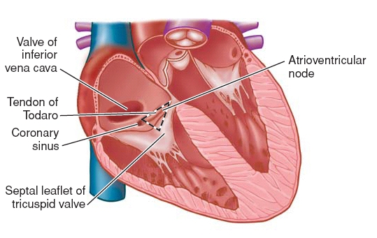
2. Atrioventricular node (AV node )
- Is situated in the right atrium , in the lower part of interatrial
septum. - It lies in the triangle of Koch, which is bounded by:
- Base of septal cusp of tricuspid valve
- Orifice of coronary sinus
- Tendon of Todaro
3. Atrioventricular bundle of HIS
- From the AV node the impulse travels in the atrioventricular bundle of HIS in the interventricular septum which divides into:
- Right ventricular branch and
- Left ventricular branch
- The two branches descend in the interventricular septum and spread out in the walls of the respective ventricles to end as Purkinje fibers.
Describe briefly the nerve supply of heart.
- The heart rate and the cardiac output are controlled by autonomic nervous system.
- Sympathetic fibers are provided by the cardiac branches (preganglionic fibers) of superior, middle and inferior cervical ganglia (preganglionic fibers reach from T2-T5 spinal segments).
- Parasympathetic fibers are provided by the cardiac branches (superior, inferior and recurrent) of the left & right vagus nerves.
The sympathetic and parasympathetic fibers reach heart via the superficial and deep cardiac plexuses.
Superficial cardiac plexus is located below the arch of aorta. it is formed by:
- Cardiac branch of superior cervical ganglion of left sympathetic chain.
- Inferior cervical cardiac branch of left vagus.
Deep cardiac plexus is located behind the arch of aorta and in front of tracheal bifurcation. It is formed by:
- Cardiac branches of middle and inferior cervical ganglion of both the sympathetic chain and from the superior cervical ganglion of right sympathetic chain.
- Cardiac branches of T2-T5 ganglion of both the sympathetic chain.
- Superior and recurrent branches of both right and left vagus nerves and inferior cardiac branch of right vagus.

