Describe the location, shape and gross features of urinary bladder.
Urinary bladder is a hollow muscular viscus that acts as a reservoir of urine.
Location
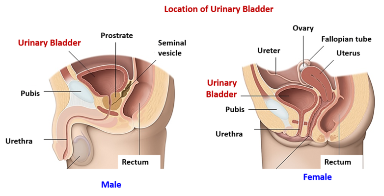
• In adults: Empty bladder lies in the pelvic cavity and the distended bladder rises above the pubic symphysis and reaches the hypogastric region of abdominal cavity.
• In infants: Urinary bladder is located in the abdominal cavity.
Shape: It is tetrahedral in shape when empty and ovoid when distended .
Gross features and relations
Empty urinary bladder presents the following parts:
• An apex: Lies at the level of upper border of pubic symphysis. It is connected to the umbilicus by median umbilical ligament ( remnant of urachus).
• Base/Fundus/Posterior surface: Is triangular in shape. In males it is related to ampulla of vas deferens and seminal vesicle. In females it is related to anterior vag vaginal wall.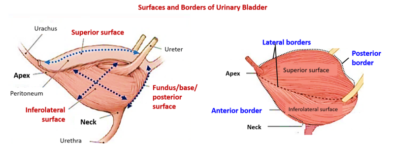
• Neck: Is the lowest and most fixed part. It lies 3-4 cm. behind the lower part of pubic symphysis. In males it is related to prostate gland & in females to pelvic fascia. It is pierced by internal urethral meatus.
Borders: There are four borders – Anterior, Posterior, and two lateral.
Surfaces:
- Superior : Is triangular in shape and is related to coils of ileum and pelvic colon.
- Two inferolateral surfaces: Are related to pubic bone, retropubic fat and levator ani.
- Posterior surface: also called base of urinary bladder.
Describe in brief the supports of urinary bladder.
Following are the Supports of urinary bladder :
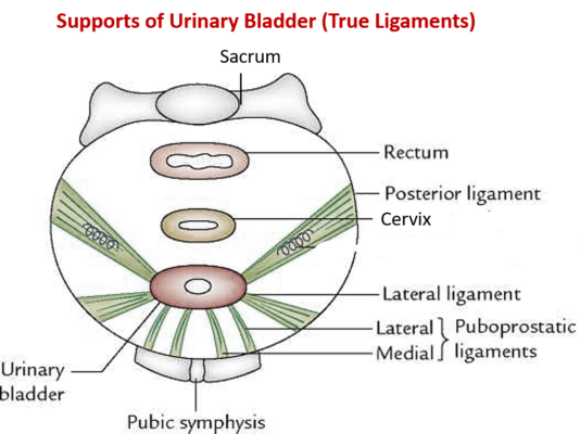
True ligaments: Are condensations of pelvic fascia.
- Lateral and medial pubovesical/puboprostatic ligaments: From posterior surface of body of pubis to the neck of the urinary bladder.
- Lateral true ligaments: From tendinous arch of levator ani muscle to inferolateral surface of the urinary bladder.
- Posterior true ligaments: base of bladder to pelvic wall along the internal iliac vessels. *
*Median umbilical ligament – from the apex of bladder to the umbilicus ( contains urachus – remnant of allantois).
False ligaments: Are formed by peritoneal folds.
- Median umbilical fold – fold of peritoneum over the median umbilical ligament.
- Lateral umbilical ligaments: from bladder to the lateral pelvic walls.
- Posterior false ligaments/sacrogenital folds: from base of bladder to sacrum.
Describe briefly the trigone of bladder.
Trigone of urinary bladder: It is an equilateral triangular area on the inner surface of base of urinary bladder.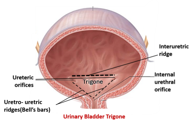
• Boundaries:
- Base – is formed by interuretric ridge (bar of Mercier) which extends between the two openings of the ureters.
- Apex – is formed by the internal urethral orifice.
- Sides – formed by uretro- uretric ridges(Bell’s bars) from the ureteric orifice to the urethral orifice
• Characteristic features:
- Mucous membrane is firmly adherent to the musculature.
- Absence of rugae (temporary folds of mucosa)
- Submucosa is absent
What is uvula vesicae?
Uvula vesicae : Is a rounded elevation just behind the urethral orifice which is formed by the median lobe of the prostate. In old aged man, the uvula vesicae may be enlarged due to enlargement of prostate and obstruct the outflow of urine through the internal urethral orifice.
Name the arteries supplying and the lymph nodes into which lymph drains from the urinary bladder.
Arteries supplying urinary bladder: It is supplied by branches of internal iliac artery. Superior vesical artery is the main arteryo. Other arteries that supply uninary bladder are obturator, inferior vesical , inferior gluteal and in females uterine artery
Lymph nodes draining urinary bladder: Internal and external iliac lymph nodes.
Describe briefly the nerve supply of urinary bladder.
Urinary bladder is supplied by autonomic nervous system.
Parasympathetic innervation: Efferent fibers arise from from S2 – S4 spinal segments and supply urinary bladder via pelvic splanchnic nerves /nervi ergentes. They are concerned with emptying of the bladder.. The fibers are motor to the detrusor muscle and inhibitory to the sphincter urethrae. The afferent fibres convey the sensation of bladder distension.
Sympathetic innervation: The efferent fibers arise from T11–L2 spinal segments and reach the urinary bladder via hypogastric plexus. The fibers are motor to sphincter urethrae muscle and inhibitory to detrusor muscle. The afferent fibers carry pain sensation. Sympathetic fibers are concerned with filling of the urinary bladder..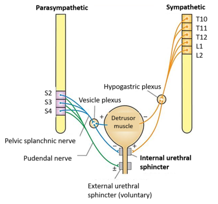
Applied Aspects
Neurogenic bladder
Dysfunction of urinary bladder due to injury/disease of nervous system (CNS/PNS) invoved in control of urination.
Automatic reflex bladder
Is an UMN (Upper motor neuron) lesion due to spinal cord lesions above T12-L1. The lesions may be due to trauma, infection, vascular infarction. There is disruption of both the sensory and motor nerve tracts above the sacral segments(S2,S3,S4). The cortical (voluntary control) is lost. The spinal reflex arc takes over control of micturition, as a result the bladder fills and empties reflexly every one to four hours.
Autonomous bladder
Occurs in LMN (Lower motor neuron) lesions, which involve disruption of the sacral spinal reflex arc at S2-S4. spinal segment level. In this case both voluntary and reflex control of micturition is lost. The bladder capacity is greatly increased due to loss of tone of deterusor muscle. The bladder overfills and overflows.

appreciate your effort. Very nicely arranged; thank you so much
Nice description & diagram.
Dr. Ferdaus
Marvellous