What are the Functions of Larynx?
Larynx is the organ of voice production (voice box) and it allows air passage as it is part of upper respiratory tract.
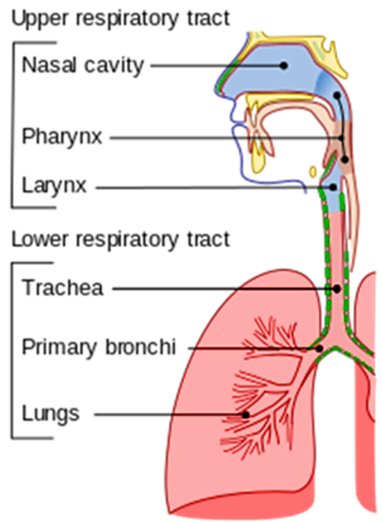
Where is larynx located and what is its Extent?
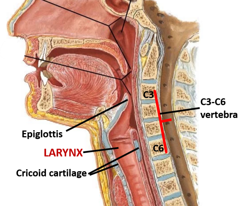
Larynx is located in front of the neck opposite 3rd to 6th cervical vertebrae in adults. It extends from the upper border of epiglottis to the lower border of cricoid cartilage.
What are the Dimensions of larynx?
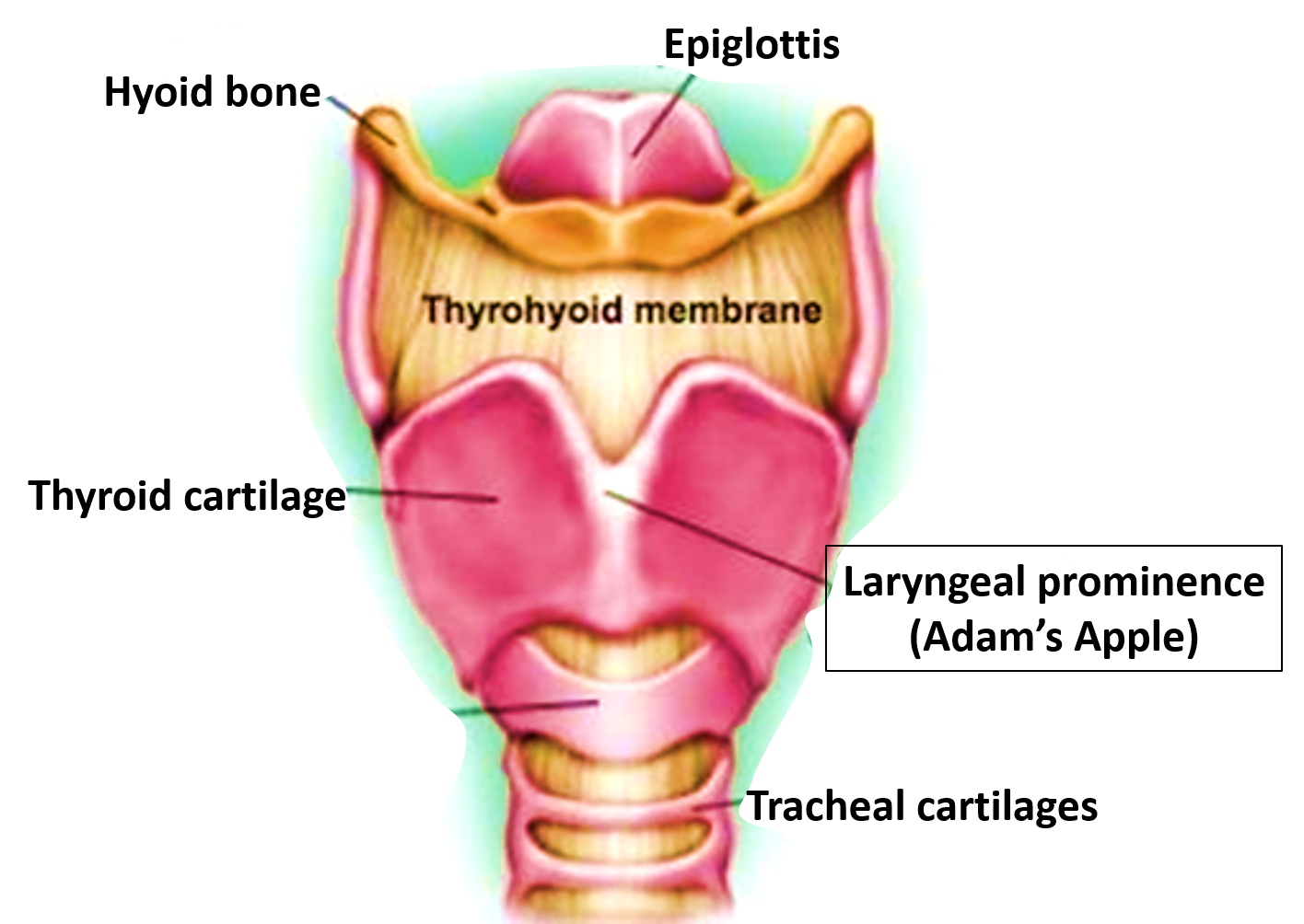
Until puberty there is little difference between the dimensions of male and female larynx. After puberty, male larynx undergoes considerable increase in size and the angle of thyroid cartilage (laryngeal prominence/ Adam’s apple) becomes prominent.
| In Males | In Females | |
|---|---|---|
| Length | 44 mm . | 36 mm . |
| Transverse diameter | 43 mm . | 4 1 mm. |
| Antero - Posterior diameter | 36 mm | 26 mm . |
| Circumference | 136 mm . | 112 mm . |
The thyroid angle (between the two laminae of thyroid cartilage) in adult males is `90o and in females it is `120o.
Name the Cartilages of Larynx?
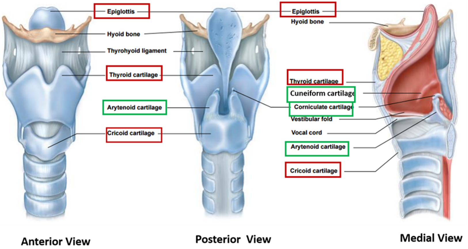
Cartilages of larynx: The framework of larynx is made up of cartilages and membranes. There are 3 paired and 3 unpaired cartilages.
Thyroid cartilage
It is a V-shaped hyaline cartilage with two laminae fused in the median plane that forms a median laryngeal prominence (Adam’s apple) particularly apparent in males. Its posterior border is continuous above with superior cornu and below with inferior cornu.
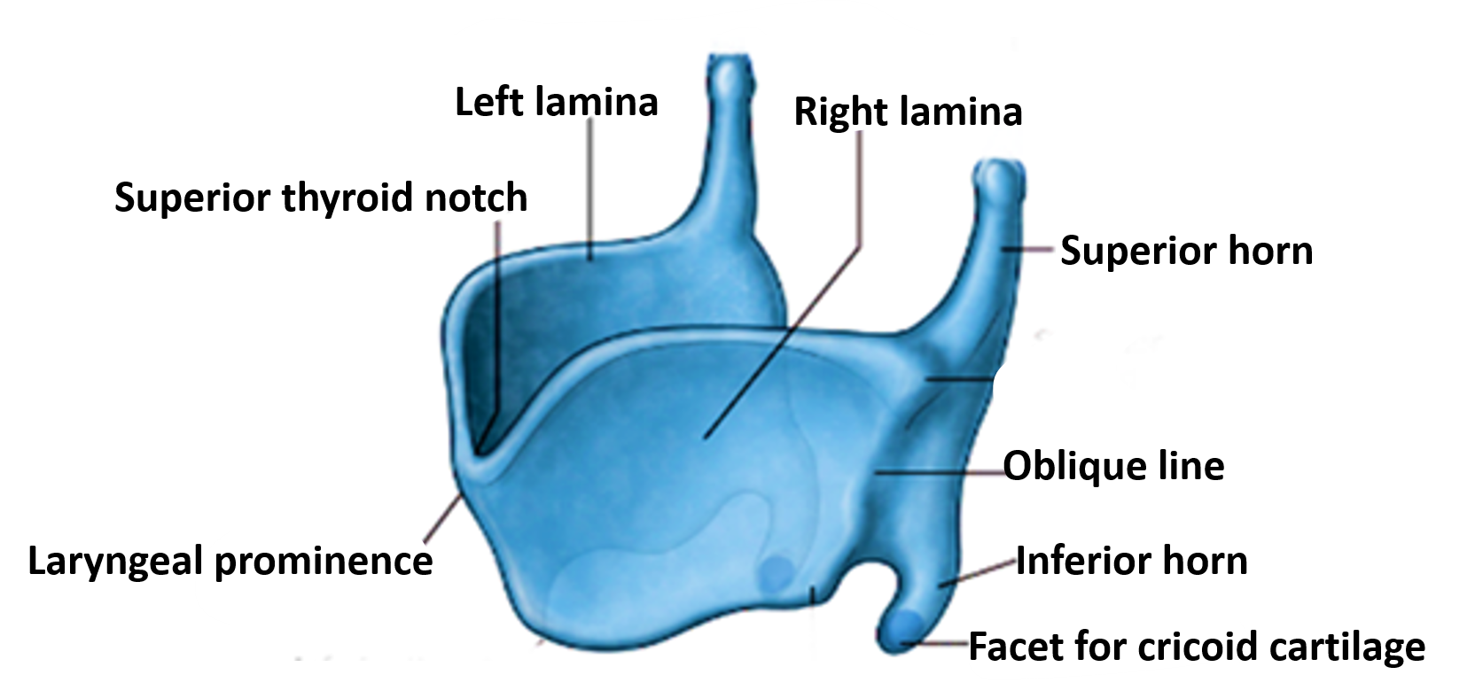
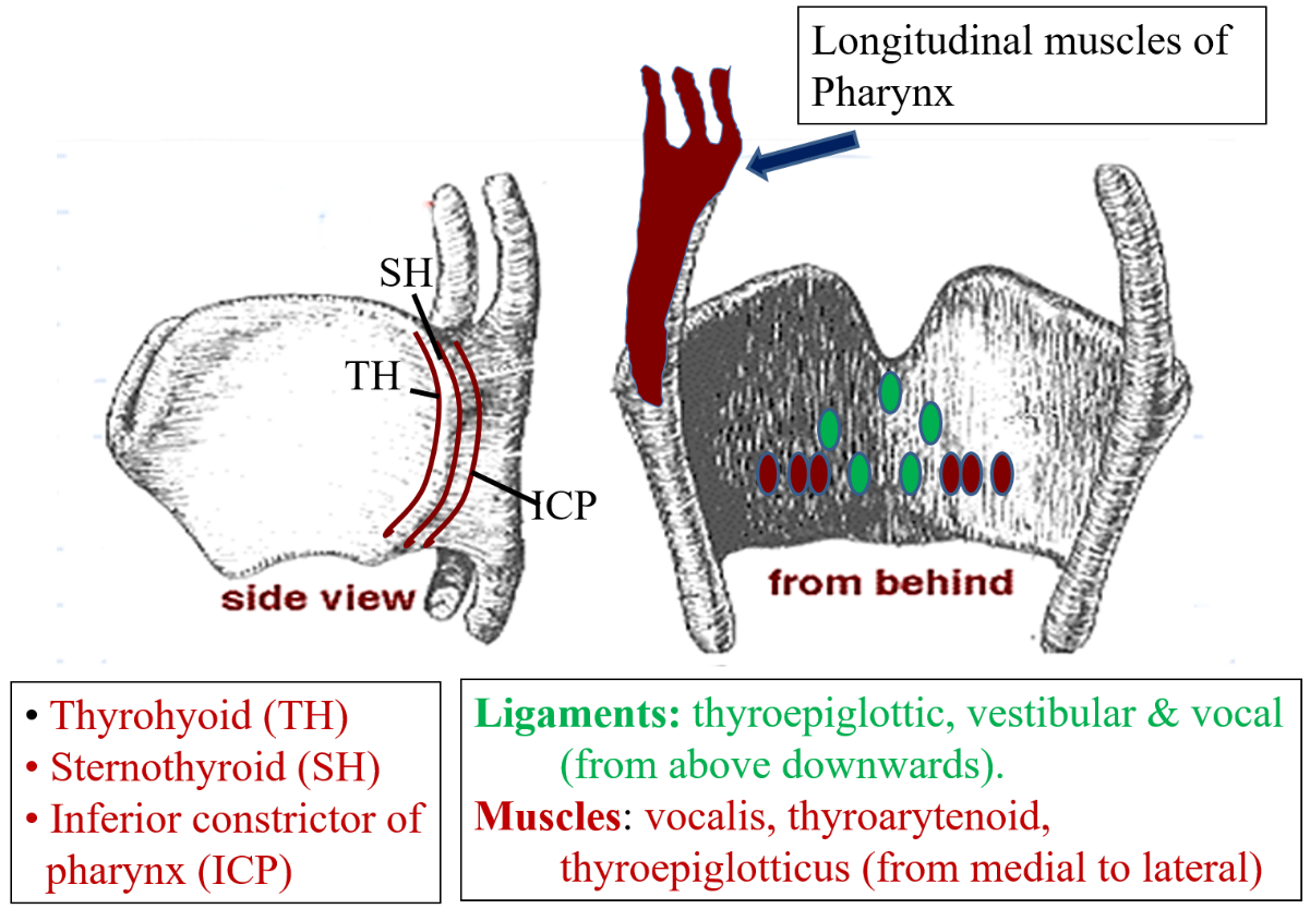
Muscles and ligaments attached to thyroid cartilage
- Oblique line on the lateral surface of lamina provides attachment to
- sternothyroid, thryrohyoid and inferior constrictor of pharynx.
- Inner surfaces of laminae near midline provide attachment to
- 5 ligaments:
- Median thyroepiglottic ligament
- Paired vestibular ligaments
- Paired vocal ligaments
- Lateral to the attachment of ligaments, it also provides attachment to 3 muscles
- Vocalis, thyroepiglottic muscle and thyroartytenoid muscle (from above down wards)).
- Posterior border provides attachment to longitudinal muscles of pharynx.
- Upper border provides attachment to thyrohyoid membrane and thyrohyoid ligaments.
- Lower border provides attachment to cricothyroid ligament.
Cricoid cartilage
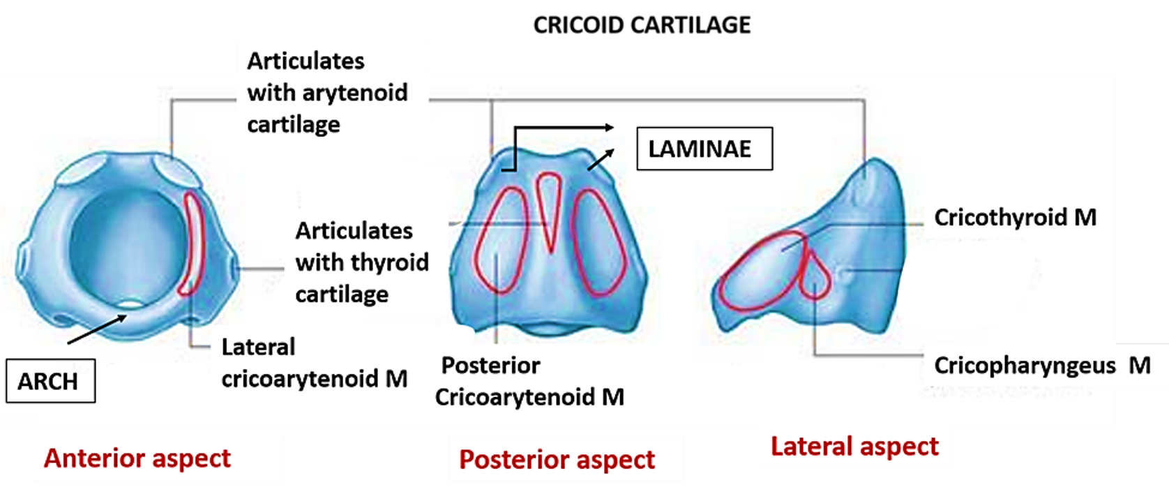
Signet ring shaped hyaline cartilage which has a narrow arch anteriorly and a wider lamina posteriorly. It lies at the level of C6 vertebra and its lower border marks the end of larynx.
Muscles attached to cricoid cartilage
- Upper border of arch – Lateral cricoarytenoid
- Posterior surface of lamina – Posterior cricoarytenoid
- Lateral surface of arch – Cricothyroid and cricopharyngeus part of inferior constrictor of pharynx.
Epiglottis
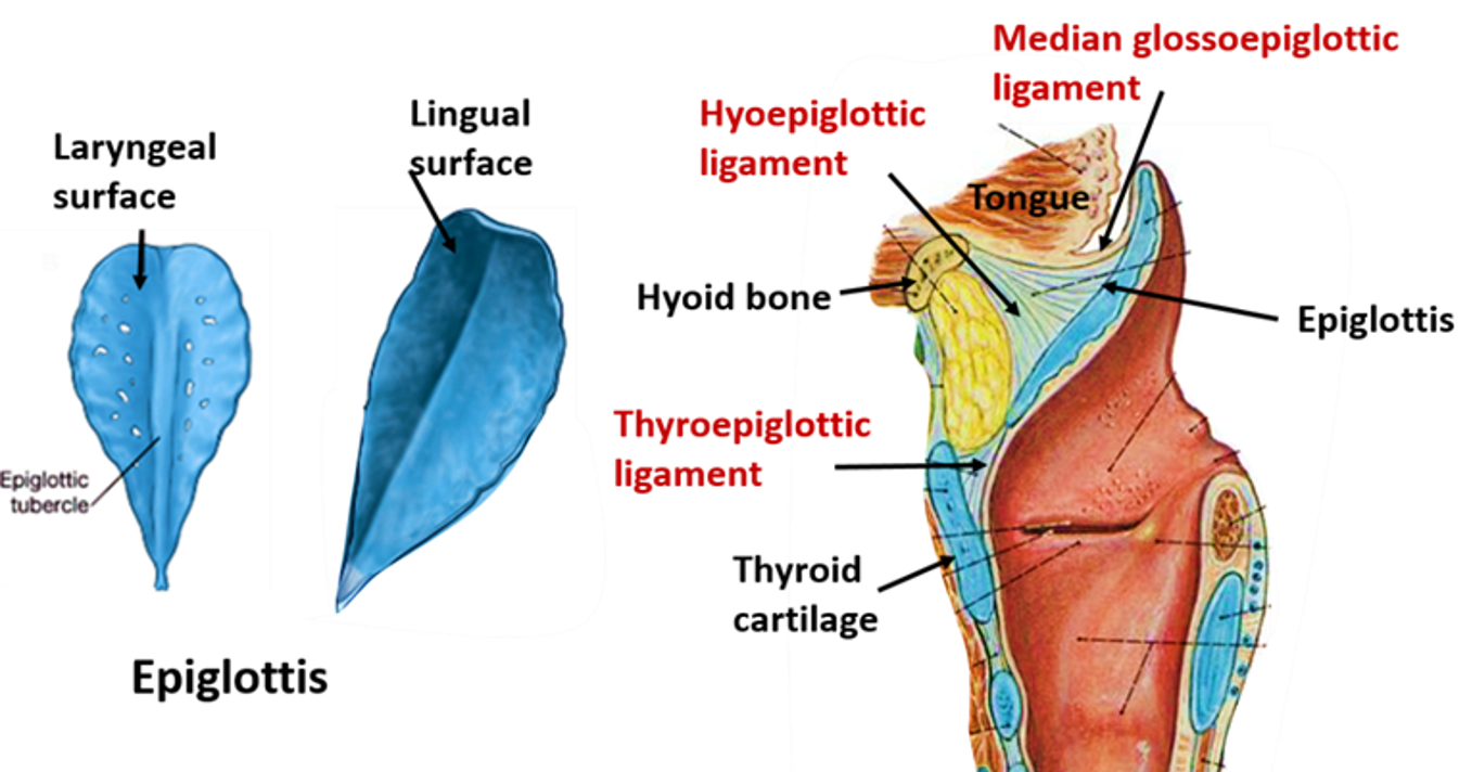
Leaf shaped elastic cartilage. It is connected anteriorly from above downwards to
- Tongue (via 2 lateral and 1 median glossoepiglottic folds)
- Hyoid bone by hyo-epiglottic ligament
- Thyroid cartilage by thyro-epiglottic ligament.
Name the Membranes and Ligaments of Larynx.
The cartilages of the larynx are interconnected to each other and to the hyoid bone and trachea by a number of ligaments and fibrous membranes.
- The extrinsic ligaments and membranes are outside the inner tube of the fibroelastic tissue of laryngeal cavity.
- The intrinsic ligaments and membranes are part of the fibroelastic tissue, present outside the mucous lining of laryngeal cavity.
| Extrinsic Ligaments | Intrinsic ligaments/membrane |
|---|---|
| • Thyrohyoid membrane | Cricovocal (conus elasticus) ligament |
| Median and lateral thyrohyoid ligaments | Quadrate/Quadrangular membrane |
| Thyroepiglottic ligament | Vocal ligament |
| Cricothyroid ligament | Vestibular ligament |
| Cricotracheal ligament |
Extrinsic ligaments/membranes
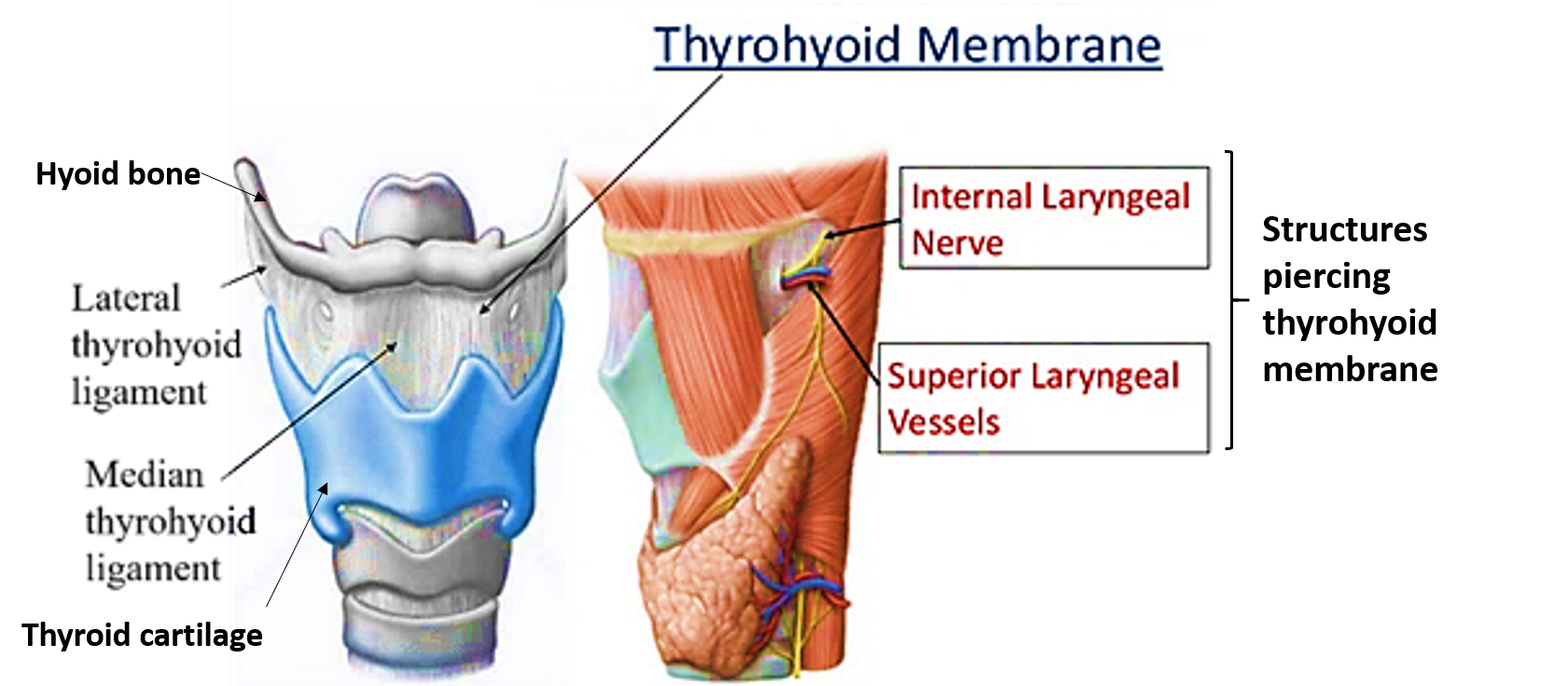
- Thyrohyoid membrane and ligaments:
- Extends between the upper border of thyroid cartilage and upper border of body and greater cornu of hyoid bone.
- Its thick median part forms median thyrohyoid ligament and thickened posterior margins form lateral thyrohyoid ligaments.
- Is pierced by internal laryngeal nerve and superior laryngeal vessels
2. Thyroepiglottic ligament: It attaches the thyroid cartilage to the epiglottis.
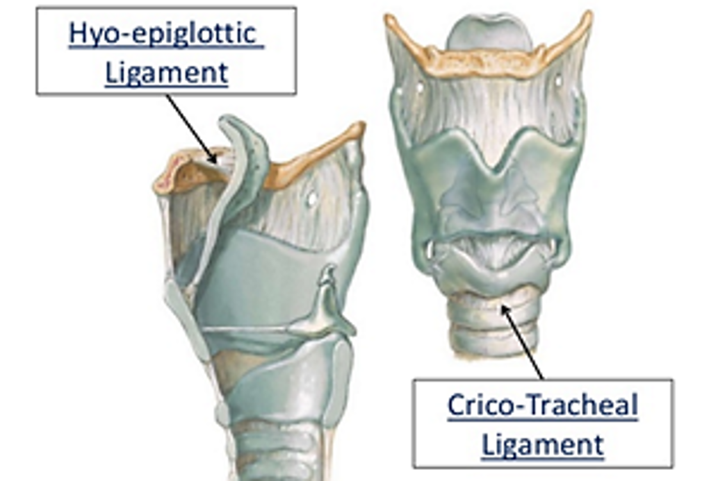
3. Cricotracheal ligament: It attaches the cricoid cartilage with the first tracheal ring.
4. Hyoepiglottic ligament: It attaches the epiglottis to the posterior surface of hyoid bone.
Intrinsic ligaments and membranes
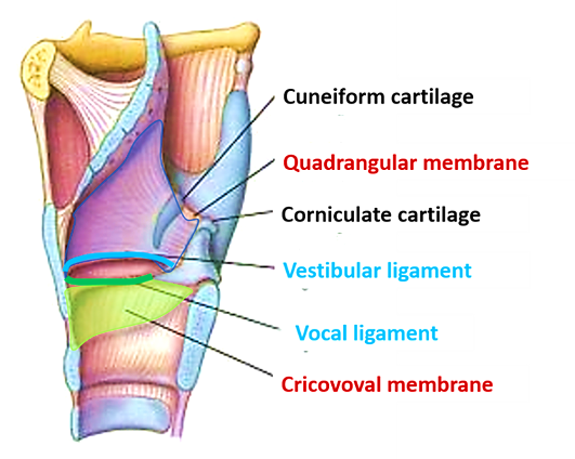
- Quadrangular/quadrate membrane:
- Is a quadrangular shaped fibroelasic membrane in the lateral wall of vestibule (supraglottic part) of larynx.
- Its upper free margin is in aryepiglottic fold and free lower thickened margin forms vestibular ligament (false vocal cord).
- Vestibular ligament is attached anteriorly to the posterior surface of thyroid cartilage and posteriorly to the lateral surface of the arytenoid cartilage
2. Cricovocal membrane/conus elasticus:
- Is a triangular fibroelastic membrane in the lateral wall of infraglottic part of the larynx.
- Its lower margin is attached to the arch of cricoid cartilage.
- Its upper thickened free margin forms vocal ligament (true vocal cord).
- Vocal ligament is attcahed antriorly to the posterior surface of the thyroid cartilage and posteriorly to the vocal process of arytenoid cartilage.
Describe Briefly the Laryngeal Cavity.
Laryngeal cavity is subdivided into three parts by two pairs of mucosal folds i.e. upper vestibular and lower vocal folds.
- Supraglottic part/Vestibule: It extends from the laryngeal inlet to the vestibular fold.
- Ventricle/sinus: It extends between the vestibular and vocal folds.
- Infraglottic part: It extends from the vocal fold to the lower border of the cricoid cartilage.
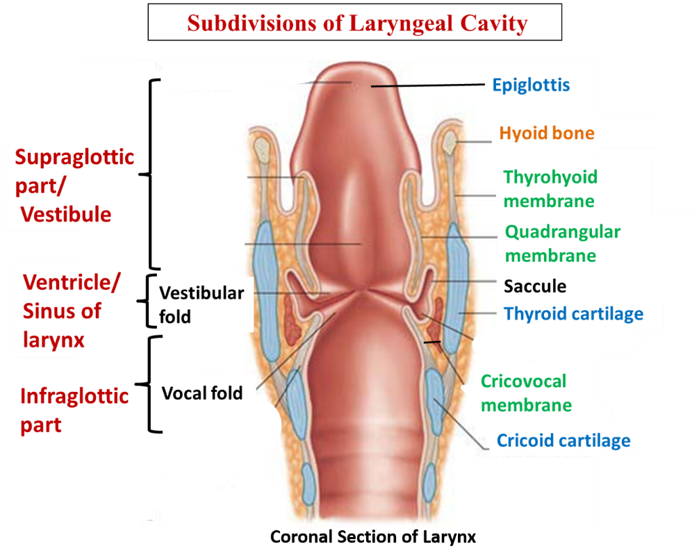
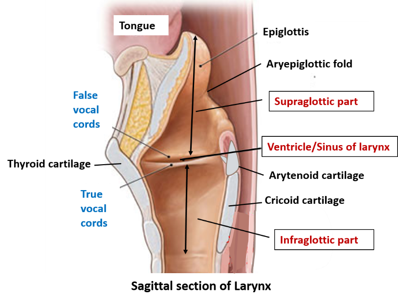
Saccule of larynx (refer above diagram): It is a blind diverticulum that extends upwards from the anterior part of sinus/ventricle of larynx between the vestibular fold and the lamina of the thyroid cartilage. It contains numerous mucous glands in the submucosal tissue. These glandular secretions of saccule keep the vocal cord moist and lubricated. Saccule is therefore known as the oil can of the larynx.
False vocal folds/vestibular folds
They are called so because they do not take part in phonation.
- Each vestibular fold contains vestibular ligament.
- They are covered by pseudostratified ciliated epithelium.
- they have submucosa.
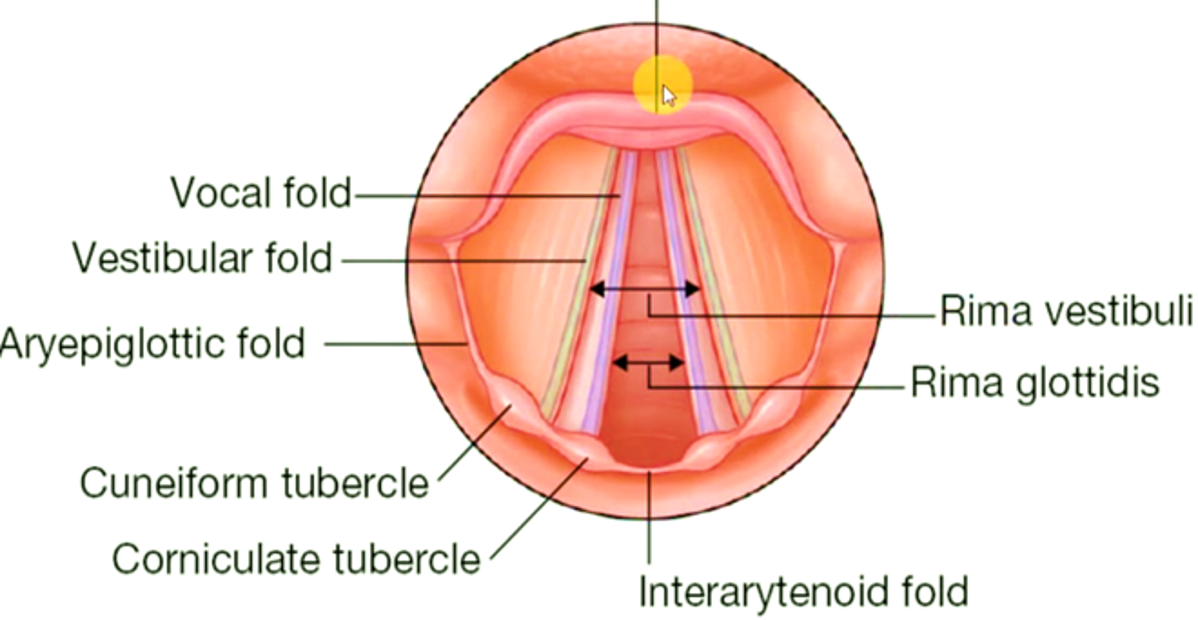
- The opening/gap between the false vocal cords is called rima vestibule.
- They appear pink in laryngoscopy due to the presence of blood capillaries in the submucosa.
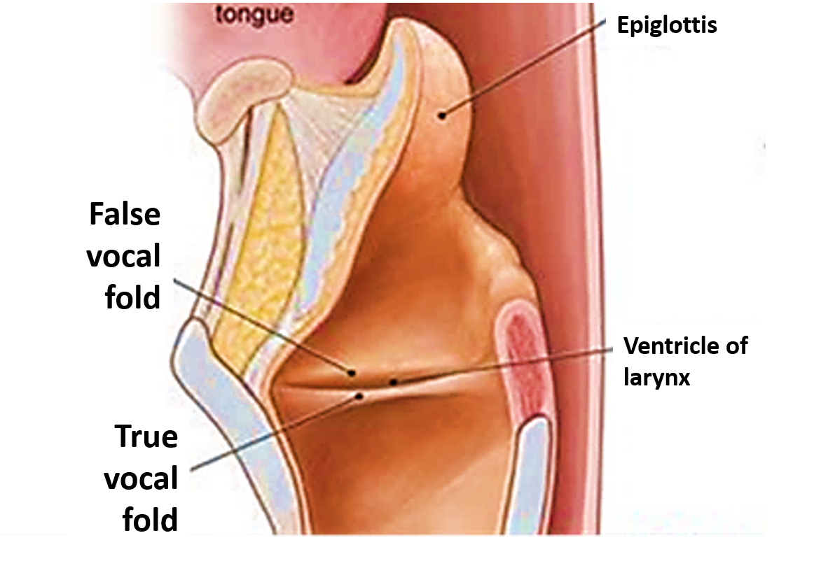
True vocal folds
They are called true vocal cords because their movement is responsible for phonation.
- Each vocal fold contains vocal ligament and vocalis muscle.
- Extend from the angle of thyroid cartilage to the vocal process of arytenoid cartilage.
- Alter the shape and size of rima glottidis during respiration and phonation.
- Are lined by stratified squamous non- keratinized epithelium.
- Appear pearly white in laryngoscopy due to the absence of blood capillaries in submucosa.
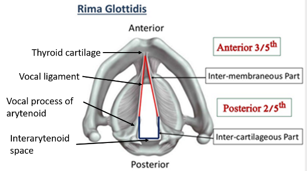
*Rima glottis is the narrowest part of the laryngeal cavity. It is bounded by:
- Vocal folds and vocal processes of arytenoids laterally.
- Thryroid cartilage anteriorly.
- Interarytenoid mucosal fold posteriorly.
- Anterior 3/5th is inter-membranous part (between the vocal ligament)
- Posterior 2/5th is inter-cartilagenous part (between the vocal processes of arytenoid cartilages)
Name the Intrinsic Muscles of Larynx and their Actions.
They connect the laryngeal cartilages and their functions are to:
- Open or close the laryngeal inlet,
- Adduct and abduct the vocal cords and
- Tense or relax the vocal cords.
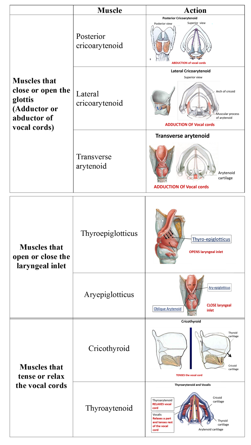
Posterior cricoarytenoid muscles are known as safety muscles of the larynx because they are the only muscles which abduct the vocal cords. If they are paralysed the unopposed action of adductors of vocal cord can block the air entry which can be fatal.
Describe the Origin, Insertion, Action and Nerve supply of Intrinsic Muscles of Larynx.
| Muscle | Origin | Insertion | Action | Nerve Supply |
|---|---|---|---|---|
| Posterior cricoarytenoid | Posterior surface of cricoid lamina lateral to median ridge | Back of muscular process of the arytenoid cartilage | Abducts vocal cord | Recurrent laryngeal nerve |
| Lateral cricoarytenoid | Upper border of cricoid arch | Front of muscular process of the arytenoid cartilage | Adducts vocal cords | Recurrent laryngeal nerve |
| Transverse arytenoid | Posterior surface of one arytenoid | Posterior surface of another arytenoid | Adducts vocal cords | Recurrent laryngeal nerve |
| Thyroepiglotticus | Posterior aspect of angle of the thyroid cartilage | Margin of epiglottis | Opens laryngeal inlet | Recurrent laryngeal nerve |
| Aryepiglotticus | Muscular process of arytenoid cartilage | Margin of epiglottis | Closes laryngeal inlet | Recurrent laryngeal nerve |
| Oblique arytenoid | Muscular process of one arytenoid cartilage | Apex of opposite arytenoid cartilage | Closes laryngeal inlet | Recurrent laryngeal nerve |
| Cricothyroid | Anterolateral part of the arch of the cricoid cartilage | Inferior cornu and adjacent part of the lower border of lamina of the thyroid cartilage | Tenses the vocal cord | External laryngeal nerve |
| Thyroarytenoid | Posterior aspect of angle of the thyroid cartilage | Anterolateral surface of the arytenoid cartilage | Relaxex vocal cord | Recurrent laryngeal nerve |
| Vocalis | Posterior aspect of angle of the thyroid cartilage | Along the vocal ligament | Relaxes a part of vocal cord and tenses the rest. | Recurrent laryngeal nerve |
Enumerate the Extrinsic Muscles of Larynx and their Actions.
They connect the larynx to the surrounding structures and are responsible for the movement of the larynx as a whole.

Describe the Nerve supply of Larynx.
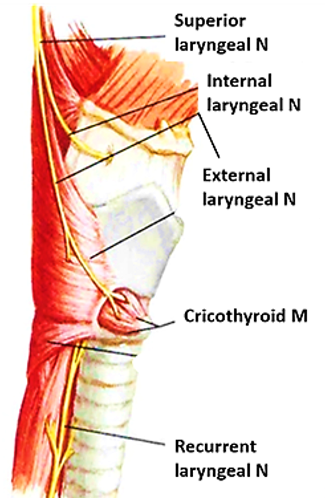
Motor supply: All the intrinsic muscles of larynx are supplied by the recurrent laryngeal nerve except cricothyroid which is supplied by external laryngeal nerve.
Sensory supply: Mucous membrane of larynx is supplied by branches of vagus nerve:
- Above the vocal cords: Internal laryngeal nerve (branch of superior laryngeal nerve).
- Below the vocal cords : Recurrent laryngeal nerve
Applied Aspects
Injury of external laryngeal nerve
If it is injured, there is weakness of phonation as a result of inability of the cricothyroid muscle to tense the vocal cords.
Injury of recurrent laryngeal nerve
May accidentally get injured during thyroidectomy:
- If injured unilaterally, the vocal cord on the affected side is located in paramedian position (between abduction and adduction) and doesn’t vibrate. But, the other vocal cord compensates and the phonation is not much affected ( hoarseness of voice).
- If injured bilaterally, both the vocal cords are placed in the paramedian position which will affect phonation and breathing.
Injury of both recurrent and external laryngeal nerves
If the recurrent and external laryngeal nerves are involved on either side, the vocal cords are farther abducted as a result of paralysis of all intrinsic muscles of the larynx. This is called the cadaveric position of vocal cords or rima glottidis. Breathing is possible but no phonation.
If internal laryngeal nerve is injured
There is anesthesia of the mucous membrane in the supraglottic part and loss of protective cough reflex. Consequently, the foreign bodies can easily goes into the larynx.
Laryngeal obstruction/choking
It is caused by aspirated food particles that are usually lodged at the rima glottidis. Choking by food can lead to asphyxia. If foreign body isn’t dislodged and expelled out instantaneously by Heimlich maneuver, it can be fatal.
Heimlich maneuver is done as follows: Stand behind the patient, pass your arms under his arms, place hands in front of his/her epigastrium with one hand make the fist and place the other hand over it. Now give three or four abdominal thrusts directed upwards and backwards. By doing this, the remaining air in the lungs is squeezed upwards in trachea and larynx with power, dislodging foreign bod.y
Laryngocele
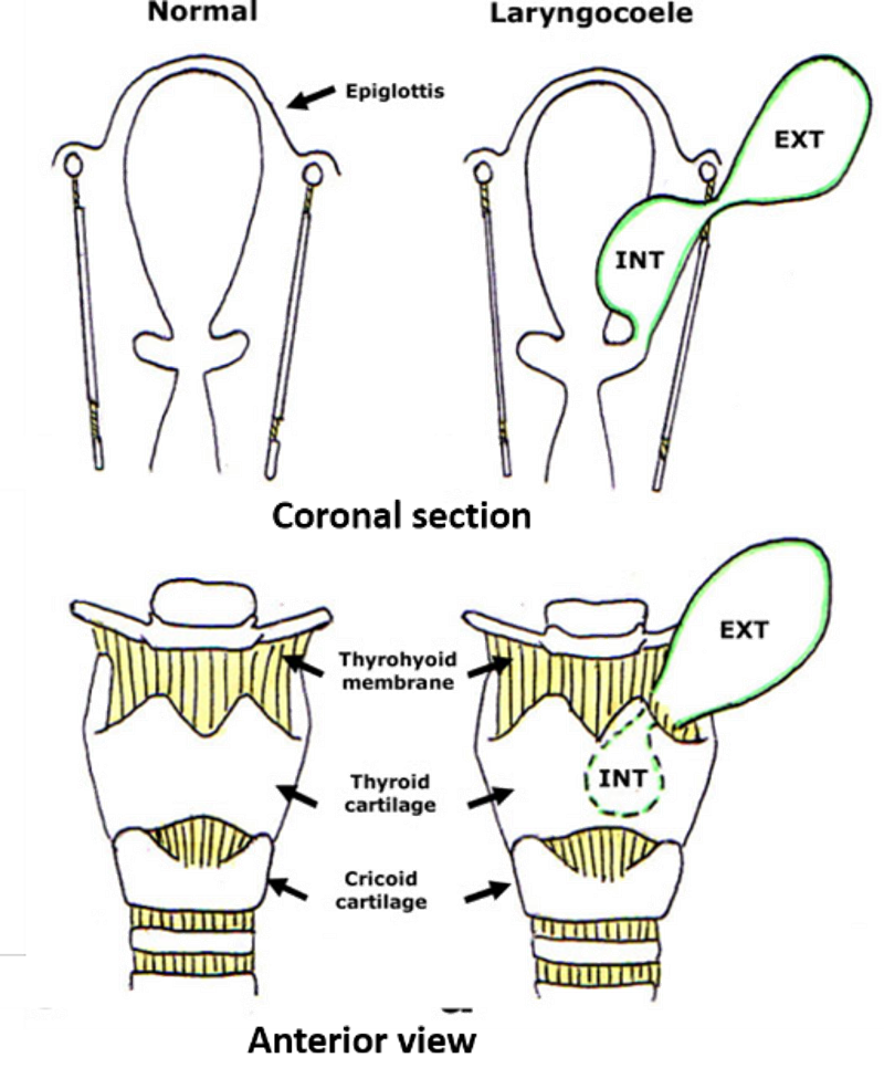
Abnormally enlarged, distended and air-filled saccule is called laryngocele. It can be congenital or acquired, as seen in, trumpet blowers and glassblowers due to continual forced expiration which increases the pressures in the larynx which leads to dilatation of the laryngeal ventricle (sinus of Morgagni) and saccule. Internal laryngocoele is limited to the larynx and confined medially by the false vocal cord, and laterally by the lamina of the thyroid cartilage. External laryngocele extends superiorly and laterally into the neck through the thyrohyoid membrane.
Vocal Nodules (Vocalist’s or Screamer’s Nodules)
During vibration of vocal cords, the point of maximum contact between the vocal cords is at the junction of their anterior one-third and posterior two-third and is subjected to maximum friction. For this reason in people, who overuse their voice, like teachers the inflammatory nodules develop at these sites named vocal nodules.

Very nice illustration Thank you
Great work
Thank you 😊