SECOND WEEK OF DEVELOPMENT
Q. By which day the blastocyst is completely embedded in the endometrium?
A. by 12th day.
Q. Name the structures derived from the inner cell mass/embryoblast.
A. Hypoblast and epiblast are formed during 2nd week of development.
Q.Name the two layers of trophoblast formed during 2nd week of development.
A. Inner layer is called Cytotophoblast (mononucleated cells) and outer layer is syncytiotrophoblast (Multinucleated cells).
Q. Name the structures derived from the trophoblast.
A. Extraembyonic mesoderm, chorion and amnion.
Q. Where is amniotic cavity located during 2nd week of development?
A. It is between the amnion above and the epiblast below.
Q. Where is extraembryonic coelom formed and what is its fate ?
A. Small cavitites which appear in the extraembryonic mesoderm join to form extraembryonic coelom which will ultimately form chorionic cavity. It splits the extraembryonic mesoderm into two layers i.e
- parietal or somatopleuric layer
- visceral or splanchnopleuric layer.
Q. What is Heuser’s membrane?
A. Flattened cells that arise from hypoblast ( according to some from the trophoblast) line the inside of blastocystic cavity form Heuser’s membrane. It encloses the primitive yolk sac.
Q. How is secondary yolk sac formed?
A. It is formed as the primary yolk sac is constricted during formation of extraembryonic coelom. Remnants of primitive yolk sac degenerate.
Q. What is chorion?
A. Extraembryonic somatopleuric mesoderm along with overlying trophoblast is called chorion.
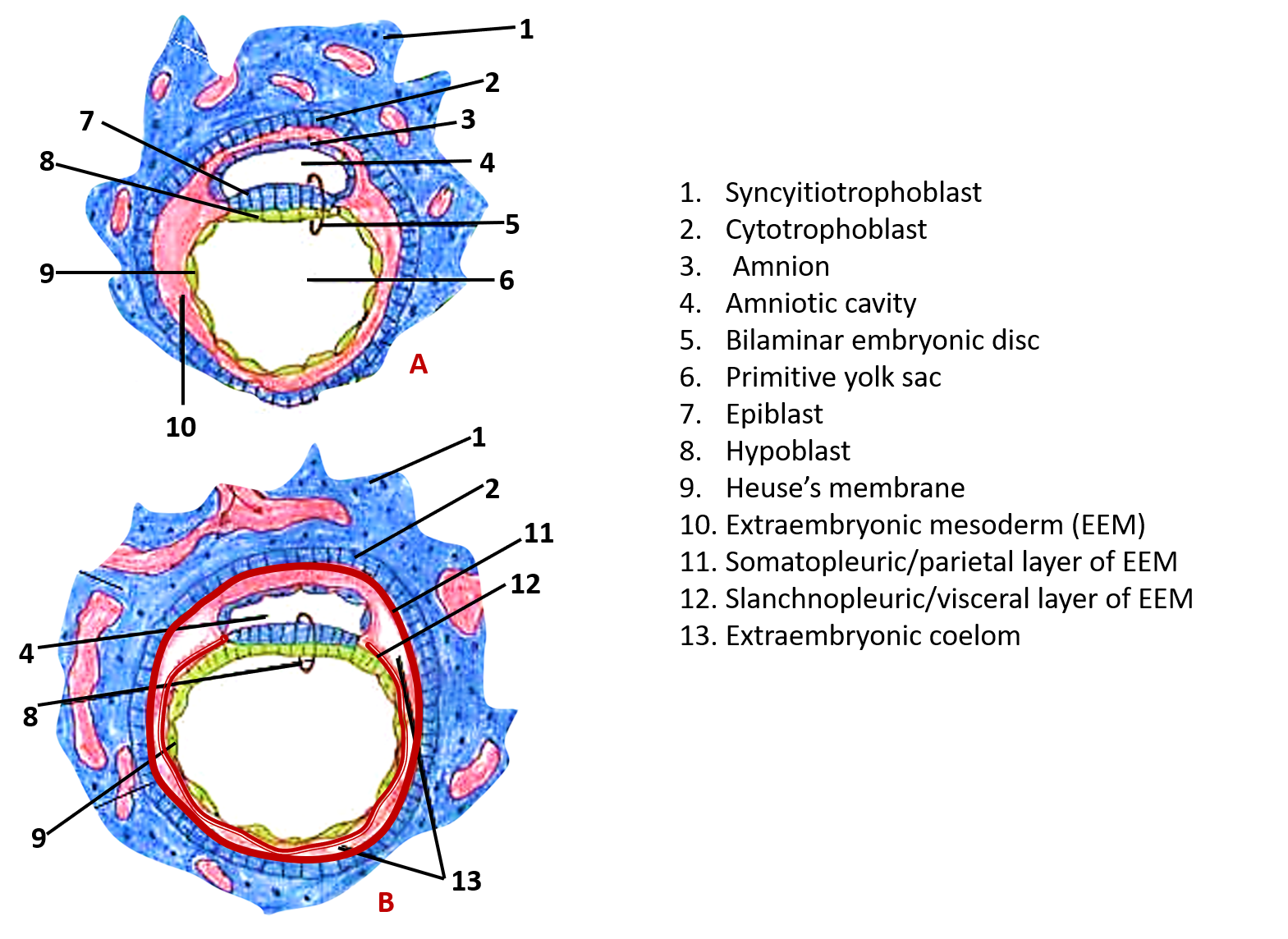
Q. Lable the structures marked 1-13.
A. 
Q. Name the structures that develop from epiblast.
A. Structures developing from epiblast are:
- Definitive endoderm
- Intraembryonic mesoderm
- Definitive ectoderm
- Notochord
- Primordial germ cells
Q. What is prochordal plate and what is its significance?
A. It is a localized circular area located at the cephalic end of embryonic disc, where endodermal cells become columnar and are firmly attached to the overlying ectoderm cells. It marks the future site of mouth. Its appearance Its appearance enable one to distinguish the future head and tail ends. It also determines the central axis of the developing embryo ( i.e. divide the embryo into right and left halves). It is formed by the end of second week of development.
Q. What is primitive streak?
A. It is a transient structure (clumps of cells) that is formed soon after the formation of prochordal plate, by the proliferation of epiblast cells along the central axis, near the tail of the embryonic disc. Its formation marks the begining of gastrulation.
Q. what is cloacal plate?
A. Localized area caudal to the primitive streak where endodermal cells are firmly attached to the ectoderm.
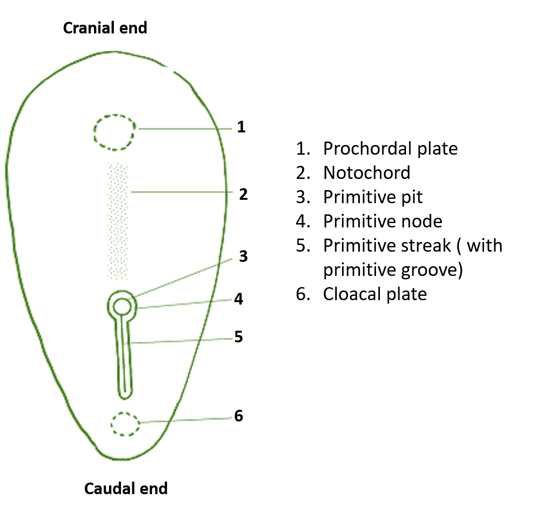
Q. Label the structures marked 1-6.

Q. What are the adult derivatives of notochord and what is its role during development?
A. Most of it disappears, but parts of it persist and form the following in adults:
- Nucleus pulposus of intervertebral disc
- Apical ligament of dens of axis (2nd) cervical vertebra.
Q. What is the role of notochord in development?
A. Notochord induces changes in the surface ectoderm to form neural plate and in surrounding mesenchyme to form paraxial mesoderm.
Q. What are the changes that occur in the 2nd week of development.
A. Following are the changes:
- Implantation is complete.
- Formation of bilaminar disc ( 2 layers i.e epiblast and hypoblast) from inner cell mass.
- Formation of 2 layers of the trophoblast ( cytotrophoblast and syncytiotrophoblast).
- Formation of amnion and chorion.
- Formation of two cavities ( amniotic and chorionic cavities).
- Formation of yolk sac.
- Formation of connecting stalk.
- Formation of prochordal plate.
- Formation of primitive streak.
Q. Name the extraembryonic membranes/structures.
A. All the structures that do not form part of embryo, but are derived from the zygote contitute extrembryonic membranes. these are:
- Chorion
- Amnion
- Yolk sac
- Umbilical cord
- Allantois

This is good way that can everyone understand embryonic lessons and i appreciated this good wey
Very nice elaborate revision material