Enumerate the muscles of the posterior/flexor compartment of leg and their nerve supply.
Muscles of this compartment are divided into three groups:superficial , middle and deep group.
- Muscles of superficial group
- Gastocnemius
- Plantaris
- Soleus
- Muscles of middle group
- Flexor digitorum longus
- Flexor hallucis longus
- Muscles of deep group
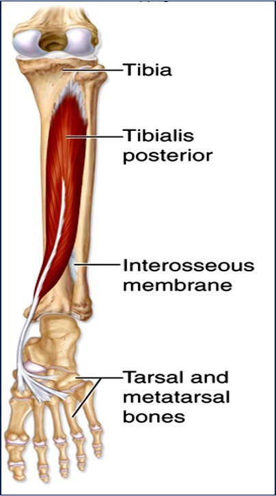
- Tibialis posterior
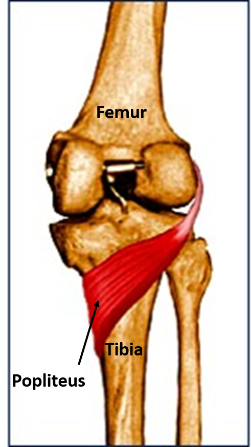
- Popliteus
* All the muscles of the posterior compartment of leg are supplied by tibial nerve.
Write the origin, insertion and action of muscles of posterior compartment of leg.
| Muscle | Origin | Insertion | Action |
|---|---|---|---|
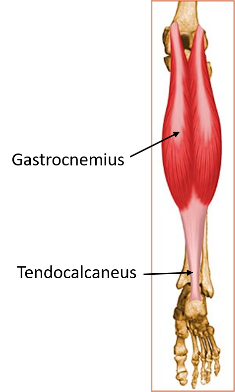
| Gastrocnemius | Lateral head: lateral condyle of femur and lower part of lateral supracondylar ridge. | Posterior surface of calcaneus via tendocalcaneus | Plantar flexion at ankle joint |
| Medial head: Popliteal surface of femur and adjoining aspect of medial condyle of femur | Flexion at knee joint | ||
| Muscle | Origin | Insertion | Action |
|---|---|---|---|
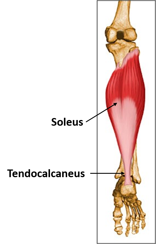
| Soleus | Upper 1/4 th of the posterior surface of shaft of fibula | Posterior surface of calcaneus via tendocalcaneus | Plantar flexion at ankle joint |
| Soleal line and middle 1/3 rd of the medial border of tibia | |||
| Muscle | Origin | Insertion | Action |
|---|---|---|---|
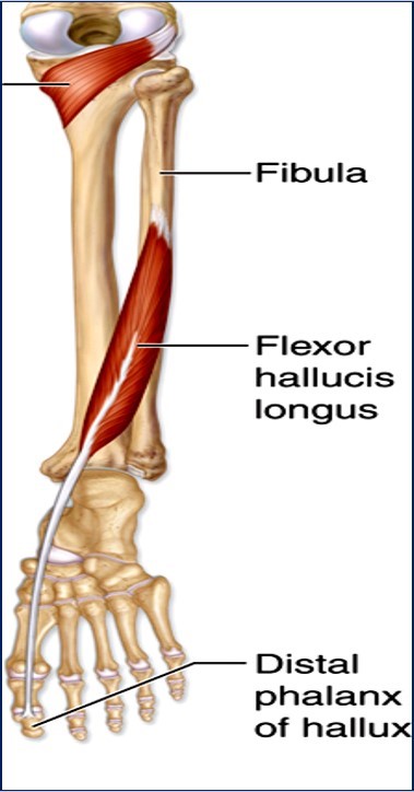
| Flexor hallucis longus | Lower 3/4 th of posterior surface of fibula between medial crest and posterior border. | Base of plantar surface of distal (terminal) phalanx of big toe. | Plantar flexion of foot at ankle joint. |
| Flexion of interphalangeal joint of big toe. | |||
| Muscle | Origin | Insertion | Action |
|---|---|---|---|
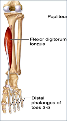
| Flexor digitorum longus | Posterior surface, medial to vertical ridge, below the soleal line. | Bases of plantar surface of distal (terminal) phalanges of lateral four toes. | Plantar flexion of foot at ankle joint. |
| Flexion of interphalangeal joints of lateral four toes. | |||
| Muscle | Origin | Insertion | Action |
|---|---|---|---|
| Popliteus | Popliteal groove on the lateral condyle of femur | Triangular area above the soleal line on the posterior surface of tibia. | Unlocks the knee joint by laterally rotating femur (foot on the ground) or medially rotating the tibia (foot off the ground) at the beginning of flexion of knee joint. |
| Muscle | Origin | Insertion | Action |
|---|---|---|---|
| Tibialis posterior | Posterior surface of tibia (below soleal line – area lateral to vertical ridge) | Chiefly on tuberosity of navicular bone | Plantar flexion of foot. |
| Posterior surface of fibula (medial to medial crest) | All tarsals except talus | Inversion of foot. | |
Enumerate the muscles responsible for plantarflexion of foot.
- Gastrocnemius
- Plantaris
- Soleus
- Tibialis posterior
- Flexor digitorum longus
- Flexor hallucis longus
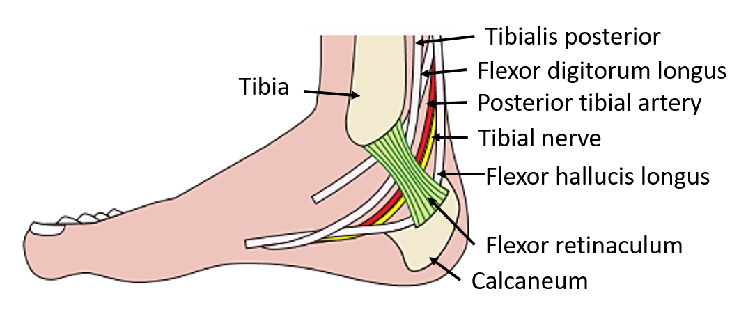
Enumerate the structures passing deep to flexor retinaculum from medial to lateral.
- Tibialis posterior
- Flexor digitorum longus
- Posterior tibial vessels
- Tibial nerve
- Flexor hallucis longus ( Mnemonic : The Doctors Are Never Hungry)
Applied Aspect.
Ruptured Tendocalcaneus
Common in middle-aged men and frequently occurs in tennis players. It occurs at its narrowest part, about 2 in. (5 cm) above its insertion. The gastrocnemius and soleus muscles retract proximally, leaving a palpable gap in the tendon. It is impossible for the patient to actively plantar flex the foot.
Intermittant Claudication
Intermittant Claudication (derived from word Claudicare (Latin) means ‘to limp’): Occurs due to ischemia of the muscles of lower limb chiefly the calf muscles. Is seen in occlusive peripheral arterial diseases particularly involving popliteal artery and its branches. The pain usually causes the person to limp. Pain is typically felt while walking and subsides with rest.

I am really pleased to say it’s an interesting post to read.
Best regards,
Abildgaard Dencker
I’m impressed