What is the Extent of Pharynx ? What are its subdivisions?
Pharynx is a funnel-shaped fibromuscular tube forming the upper part of air and food passage. It is 12-14 cm. long. Its width at upper end is 3.5cm. and at lower end 1.5 cm. It is continuous inferiorly with esophagus.
- Extent: From base of skull to the lower border of cricoid cartilage (vertebral level C6).
- Subdivisions: The anterior wall of pharynx is deficient anteriorly as it opens into nasal cavity, oral cavity and larynx from above downwards. Therefore, the subdivisions of pharynx are named:
- Nasopharynx
- Oropharynx
- Laryngopharynx
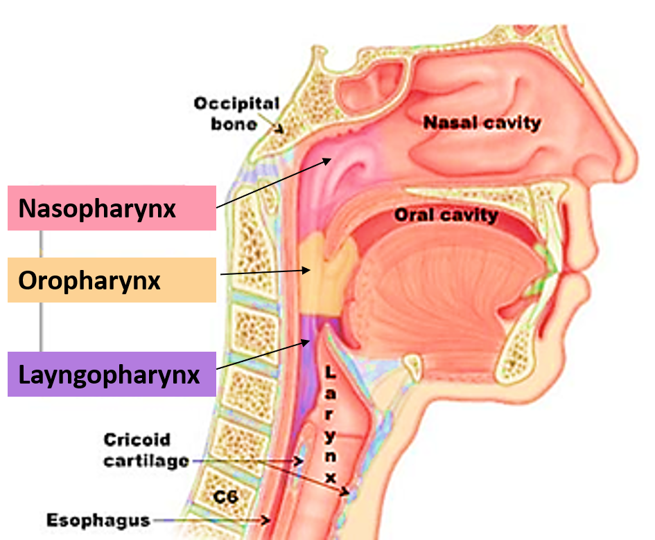
Describe the Features of Nasopharynx, Oropharynx and Laryngopharynx.
Nasopharynx
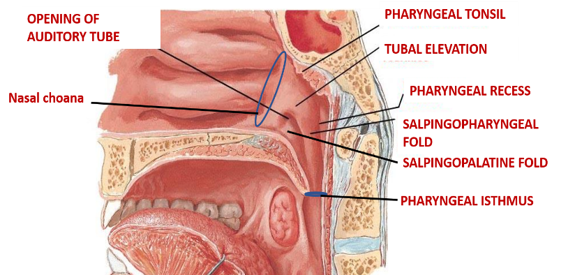
It extends from base of skull to the soft palate.
- It communicates:
- anteriorly with nasal cavity through nasal choanae.
- inferiorly with oropharynx through pharyngeal isthmus
- laterally thorough auditory tube with middle ear.
- Pharyngeal tonsil projects in the nasopharynx from the junction of its roof and posterior wall.
- The medial end of auditory/ pharyngotympanic tube opens through tubal orifice into the lateral wall on each side.
- Tubal orifice is bounded above and posteriorly by tubal elevation. Tubal tonsil is located here.
- Two mucosal folds extend from the tubal elevation.
- Salpingopalatine fold containing levator palatini muscle.
- Salpingopharyngeal fold containing salpingopharyngeus muscle.
Oropharynx
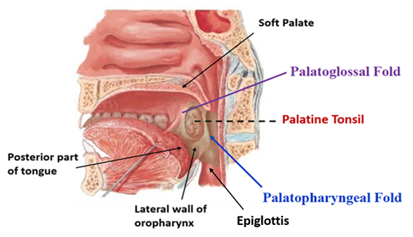
It extends from soft palate to the tip of epiglottis.
- It Communicates:
- Above : with nasopharynx through pharyngeal isthmus.
- Below : with laryngopharynx.
- In front : with oral cavity through oropharyngeal isthmus (between the palatoglossal arches)
- Lateral wall presents palatoglossal and palatopharyngeal arches enclosing tonsillar fossa for the palatine tonsil.
Laryngopharynx
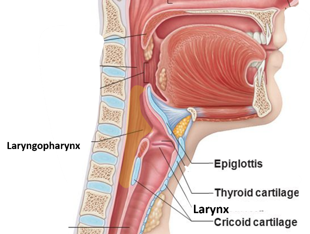
It extends from upper border of epiglottis to the lower border of cricoid cartilage.
- It communicates:
- Above: with oropharynx
- Below: with oesophagus
- Anteriorly: with larynx through laryngeal inlet.
Pyriform fossa
- Lateral wall of laryngopharynx has recess known as piriform fossa on either side of the laryngeal inlet.
- The boundaries of piriform fossa are:
- Medial: aryepiglottic fold
- Lateral: mucosa covering the thyrohyoid membrane and lamina of thyroid cartilage.
- One important feature of piriform fossa is that internal laryngeal nerve and superior laryngeal vessels pierce the thyrohyoid membrane and traverse from lateral to medial aspect beneath the mucosa of the floor of piriform fossa to reach larynx (for applied aspect see below).
Enumerate the Layers of wall of Pharynx.
Layers of pharyngeal wall from inside out are:
- Mucosa
- Submucosa
- Pharyngobasilar fascia
- Muscular coat inner longitudinal and outer circular muscles)
- Buccopharyngeal fascia
Enumerate the Muscles of Pharynx and Write their Nerve Supply.
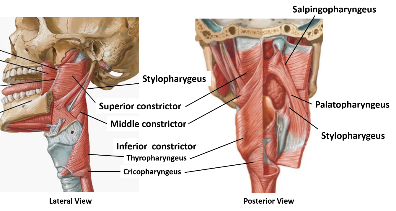
Muscles of pharynx: Pharynx has 3 circular and 3 longitudinal muscles.
Circular muscles: Superior, middle and inferior constrictors.
Longitudinal muscles: Stylopharyngeus, palatopharyngeus and salpingopharyngeus.
All the muscle of pharynx are supplied by vago-accessory complex via pharyngeal plexus of nerves except stylopharyngeus which is supplied by glossopharyngeal nerve.
Which Nerves Provide Sensory Innervation to Pharynx?
Sensory nerves supplying pharynx
- Nasopharynx: by pharyngeal branch of pterygopalatine ganglion (fibers from maxillary nerve).
- Oropharynx: by glossopharyngeal nerve
- Laryngopharynx: by internal laryngeal nerve (branch of superior laryngeal, which is a branch of vagus nerve).
Enumerate the Structures Passing Between the Constrictors of Pharynx
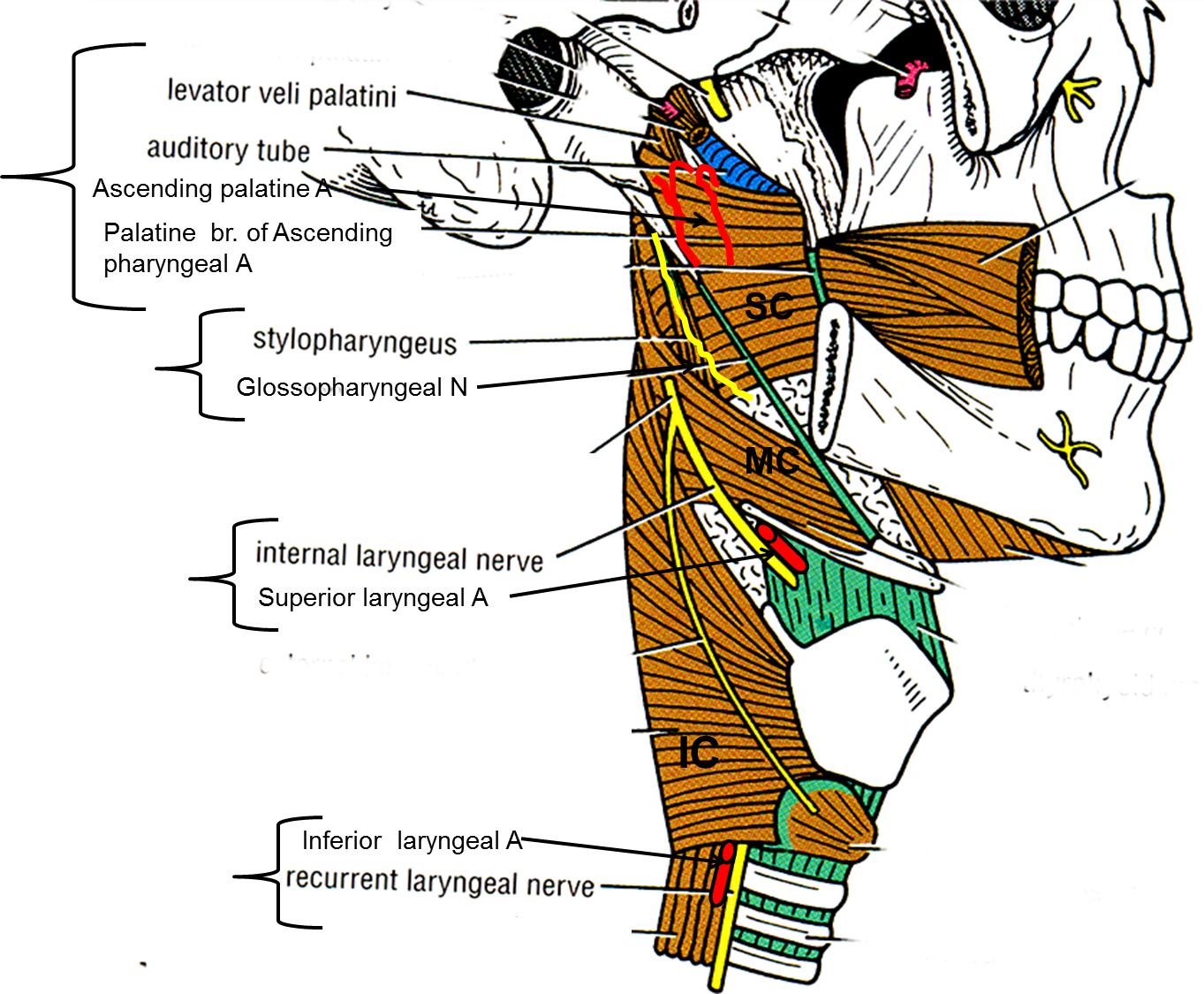
Structures passing above the superior constrictor
- Auditory tube
- Levator veli palatini
- Ascending palatine artery
- Palatine branch of ascending pharyngeal artery
Structures passing between superior and middle constrictor
- Stylopharyngeus muscle
- Glossopharyngeal nerve
Structures passing between middle and inferior constrictor
- Internal laryngeal nerve
- Superior laryngeal vessels
Structures passing below the inferior constrictor
- Recurrent laryngeal nerve
- Inferior laryngeal vessels
What is Passavant’s Ridge?
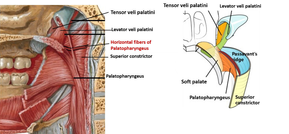
Passavant’s ridge: Some fibers of palatopharyngeus muscle pass horizontally backwards from the palatine aponeurosis along the lateral and posterior wall of nasopharyngeal isthmus to blend with superior constrictor muscle. When the soft palate is elevated the muscle forms a mucosal ridge (Pasavant’s ridge). During swallowing the soft palate and Pasavant’s ridge are approximated to shut off nasopharynx from oropharynx which prevents regurgitation of food into the naopharynx and nasal cavity.
Killian’s Dehiscence and Pharyngeal Diverticulum
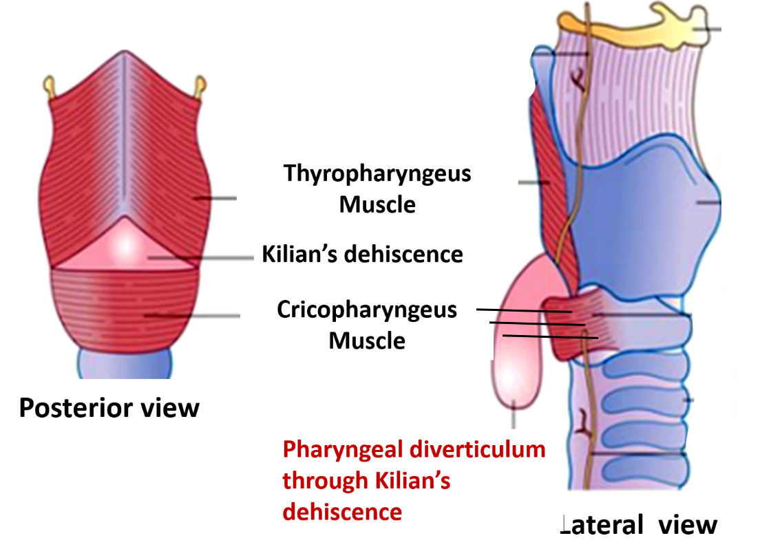
Inferior constrictor muscle has two parts viz. thyropharyngeus and cricopharyngeus. Fibers of thyropharyngeus pass obliquely to the posterior median raphe, whereas the fibers of cricopharyngeus run horizontally.A potential gap therefore exists between the thyropharyngeus and cricopharyngeus muscles which is seen as a dimple in the lining mucosa of this region of pharynx called Killian dehiscence .
Pharyngeal diverticulum may develop through this weak area of pharynx in case of neuromuscular incoordination i.e. if cricopharyngeus fails to relax following contraction of thyropharyneus part. The increased pressure forces the mucosa and submucosa posteriorly through the Killian dehiscence. The diverticulum gradually enlarges in downward direction behind the pharynx.
Applied Aspect
Adenoids
Hypertrophy of pharyngeal tonsil is called adenoids.
Pharyngeal bursa
A midline pharyngeal bursa present in the pharyngeal tonsil denotes the site of attachment of notochord to the endoderm of primitive pharynx.
Pharyngeal hypophysis
A depression in the mucosa above the pharyngeal tonsil denotes the site of path of Rathke’s pouch. Sometimes remnant cells of Rathke’s pouch may give rise to pharyngeal hypophysis, which may become malignant.
Injury to internal laryngeal nerve in pyriform fossa
Occasionally ingested food particles such as fish bone may get impacted in the fossa. If care is not taken while removing the foreign particle, internal laryngeal nerve may be injured causing loss of sensations from the supraglottic part of larynx which leads to loss of protective cough reflex.
Gag Reflex
Touching soft palate or posterior third of tongue results in:
- Reflex contraction of pharyngeal muscles
- Elevation of soft palate
- Retching and finally vomitting may be triggered.
This is known as ‘Gag Reflex’. It prevents something from entering pharynx except as part of normal swallowing. Glossopharyngeal nerve carries the afferent fibers for the reflex and Vagus nerve carries the efferent fibers.

Good day
How do I access the rest of the information on short and long questions, diagrams, anatomical basis. I am unable to access it.
Thank you