Advertisements
What is the Extent and Parts of Ureter?
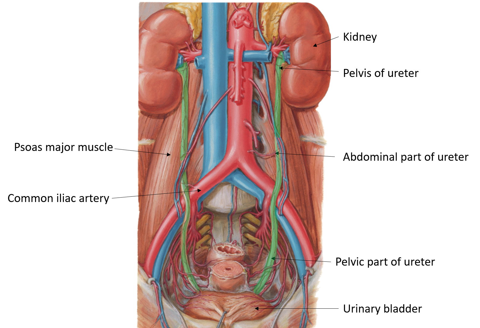
The Ureters are a pair of thick-walled, narrow muscular tubes that drains the urine from the kidney to the urinary bladder by peristaltic contractions of the smooth muscle in their wall.
Each ureter is about 25 cm /10 inches long and has a diameter of approximately 3mm.
Ureter is divided into 3 parts:
- Pelvis of ureter.
- Abdominal part (extends from the lower end of kidney till the bifurcation of common iliac vessels/sacroiliac joint/pelvic brim).
- Pelvic part (extends from bifurcation of common iliac vessels/sacroiliac joint/pelvic brim till base of the bladder).

Describe briefly the course of ureter.
- The pelvis of ureter (capacity is 5-7ml) is a funnel shaped dilatation at the upper end of the ureter which is formed within the renal sinus by joining of major calyces. It leaves the kidney by passing through the hilum of kidney (posterior to the renal vein and renal artery) and continues downward as abdominal part of the ureter at the lower pole of the kidney.
- The abdominal part of the ureter passes downwards and slightly medially deep to the peritoneum of posterior abdominal wall on the anterior surface of Psoas major muscle. The psoas major muscle separates it from the transverse processes of lumbar vertebrae.
- Pelvic part of the ureter enters the pelvic cavity by passing in front of the bifurcation of common iliac artery, in front of the sacroiliac joint. In the pelvis, the ureter first runs downward, backward, and laterally along the anterior margin of the greater sciatic notch and reaches the level of ischial spine. From the ischial spine, it turns forwards and medially to reach the superolateral angle of the base of urinary bladder, where it enters the bladder wall. It passes obliquely in the submucosa of the urinary bladder and opens into the cavity of the bladder at the lateral angle of the trigone. The ureteric openings are about 2.5 cm apart in empty bladder.
Name the sites of constrictions of Ureter.
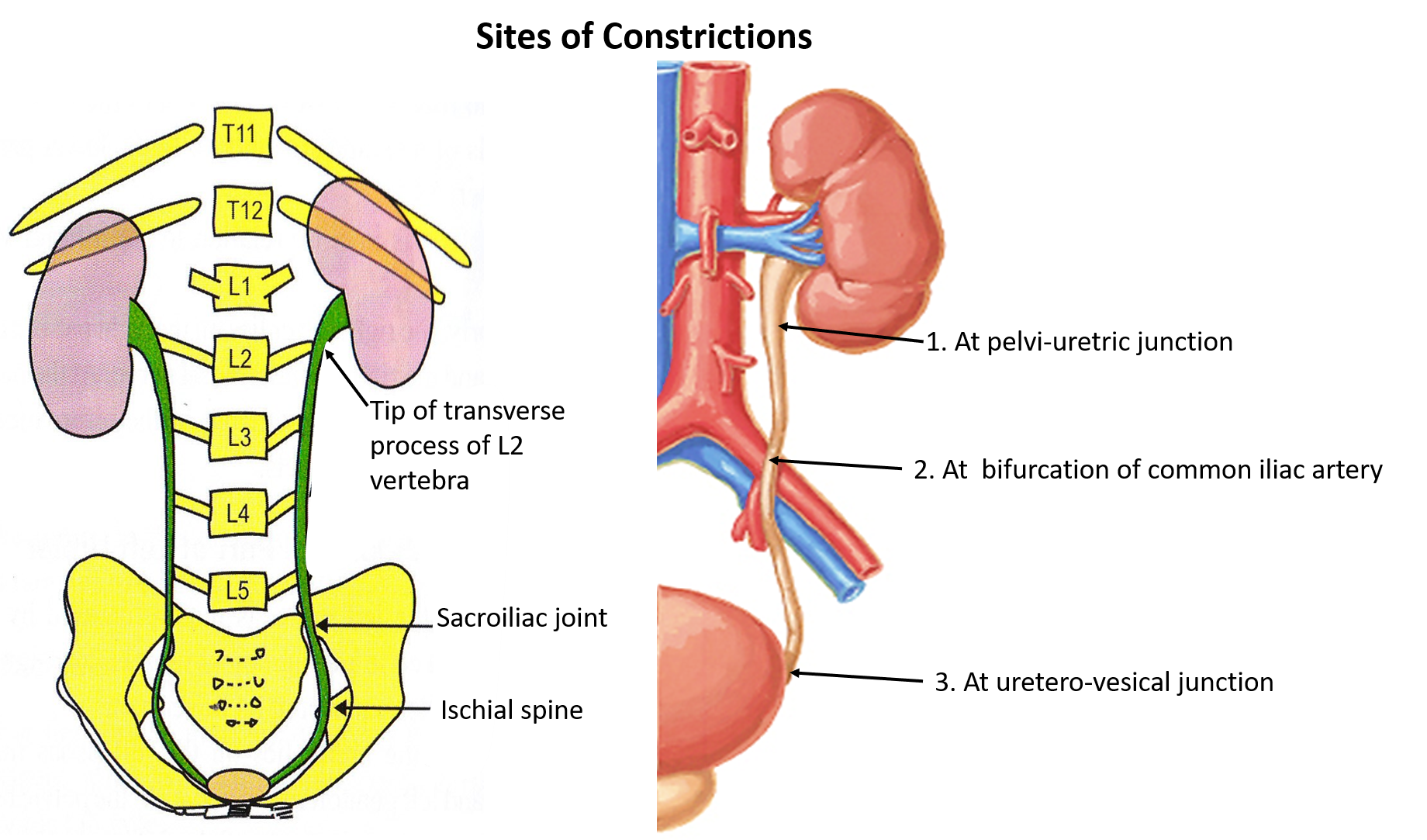
Usually there are three constrictions which are present at the following locations:
- At the pelvi-uretric junction which is at the level of lower pole of kidney (at the level of tip of transverse process of 2nd lumbar vertebra).
- At the pelvic brim (at sacroiliac joint).
- At uretero-vesical junction, the point where ureter pierces the urinary bladder (at the level of ischial spine).

Name the arteries that supply Ureter.
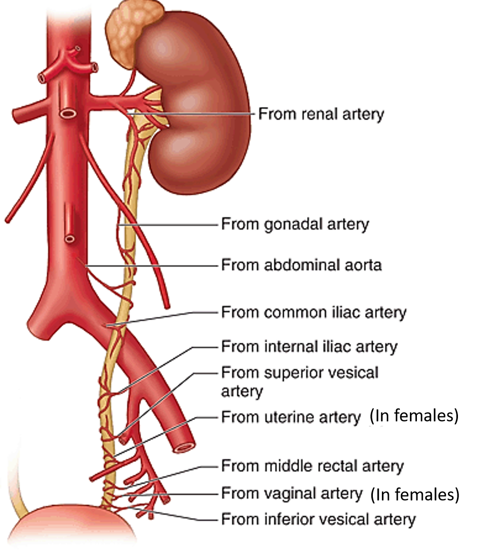
Ureter is supplied by branches of the following arteries from above downwards:
- Renal
- Gonadal (testicular or ovarian)
- Abdominal aorta
- Internal iliac
- Superior and Inferior vesical
- Middle rectal.
- Uterine (in females)
 Abdominal part of ureter is supplied by the branches from its medial side.
 Pelvic part is supplied by branches of arteries on its lateral side.
The branches from these arteries divide into ascending and descending branches and forms longitudinal anastomosis along the ureter.
Where does the lymph from Ureter drains?
- Lymph from the abdominal part drains into para-aortic lymph nodes.
- Lymph from the pelvic part drains into common iliac and internal iliac lymph nodes.
Describe in brief the nerve supply of ureters.
- Sympathetic nerve supply of the ureter is derived from T10 to L1 spinal segments which reaches ureter via renal and superior hypogastric plexuses.
- Parasympathetic nerve supply of ureter is derived from S2 to S4 spinal segments via pelvic splanchnic nerves.
Applied Aspects
Uretric Calculus
Ureteric calculus/stones are most commonly lodged at the sites of ureteric constriction i.e.
- At the pelvi-ureteric junction.
- At the pelvic brim .
- In the intramural part.
Ureteric colic
The referred pain in case of ureteric colic (due to ureteric stone) is felt in the following regions.
- In case of upper ureteral obstruction it is referred to the loin/ lumbar region (both supplied by T12 and L1 spinal segments).
- In case of middle ureteral obstruction it is is referred to the groin/inguinal and pubic regions (both supplied by L1, L2 spinal segments).
- In case of lower ureteral obstruction the pain is referred to the perineum (both supplied by S2 -S4 spinal segments).
Injury to Ureter
The ureter may get injured at the following sites
- In the infundibulopelvic ligament, during ligation of ovarian vessels as it runs very close to the ovary.
- During hysterectomy (surgical removal of uterus) ureter may be mistakenly clamped along with uterine artery. Near the cervix the ureter is crossed above by the uterine artery. The relationship between theses two structures is remembered by the phrase” water under the bridge“.

It’s ril a very interesting and understandable literature…thanks