Describe the location, dimensions and gross features of liver.
Liver is the largest gland of the body. It consists of both exocrine (secretes bile into ducts) and endocrine (secretes plasma proteins, heparin etc. into the blood) parts.
Location: it occupies whole of right hypochondrium, upper part of epigastrium and left hypochondrium (upto left midclavicular line).
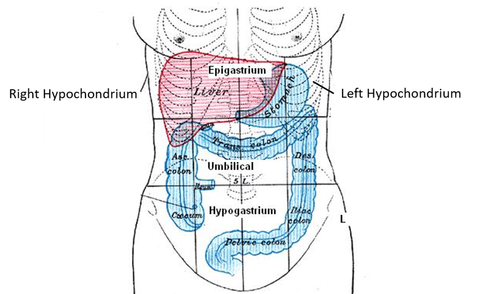
Shape: Wedge shaped.
Weight: average 1.5 kg (ratio of body weight: weight of liver – in adults: 1/36 , in newborn: 1/18. Liver is large in newborn due to the haemopoietic activity)
Gross features: Liver has 5 surfaces: anterior, posterior, superior, inferior, right lateral. Only one prominent border i.e. inferior border.
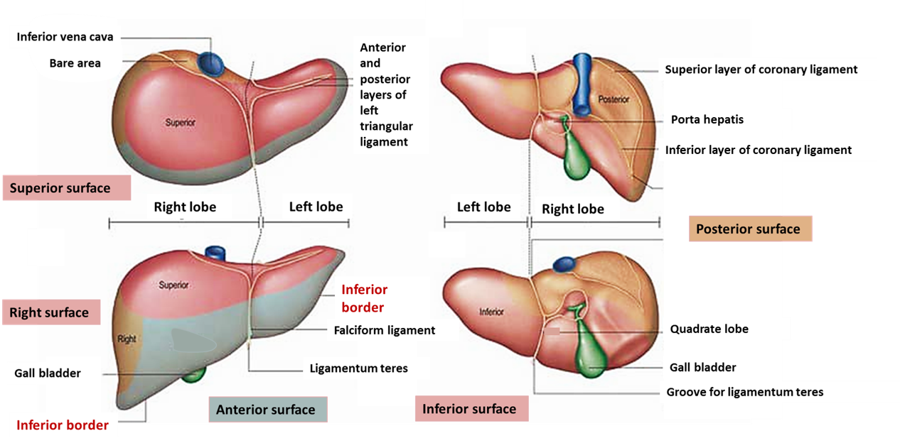
Describe the anatomical and physiological/surgical lobes of liver(see the diagram above).
Anatomical Lobes: The liver is divided into two lobes viz. right and left.
Demarcation:
- Anteriorly and Superiorly: attachment of falciform ligament.
- Inferiorly: fissure for ligamentum teres hepatis.
- Posteriorly: fissure for ligamentum venosum.
Physiological lobes: The liver is divided into right and left lobes on the basis of distribution of hepatic artery, portal vein and hepatic duct (i.e. right lobe receives blood from the right branches of hepatic artery and portal vein and pours bile into right hepatic duct)
Demarcation:
- Anterosuperiorly: the plane passes along the line joining the cystic notch to the groove for inferior vena cava.
- Inferiorly: through fossa for gall bladder.
- Posteriorly: through the middle of the caudate lobe.
Enumerate the bare areas of liver.
Bare areas of liver are the areas that are not covered by peritoneum.
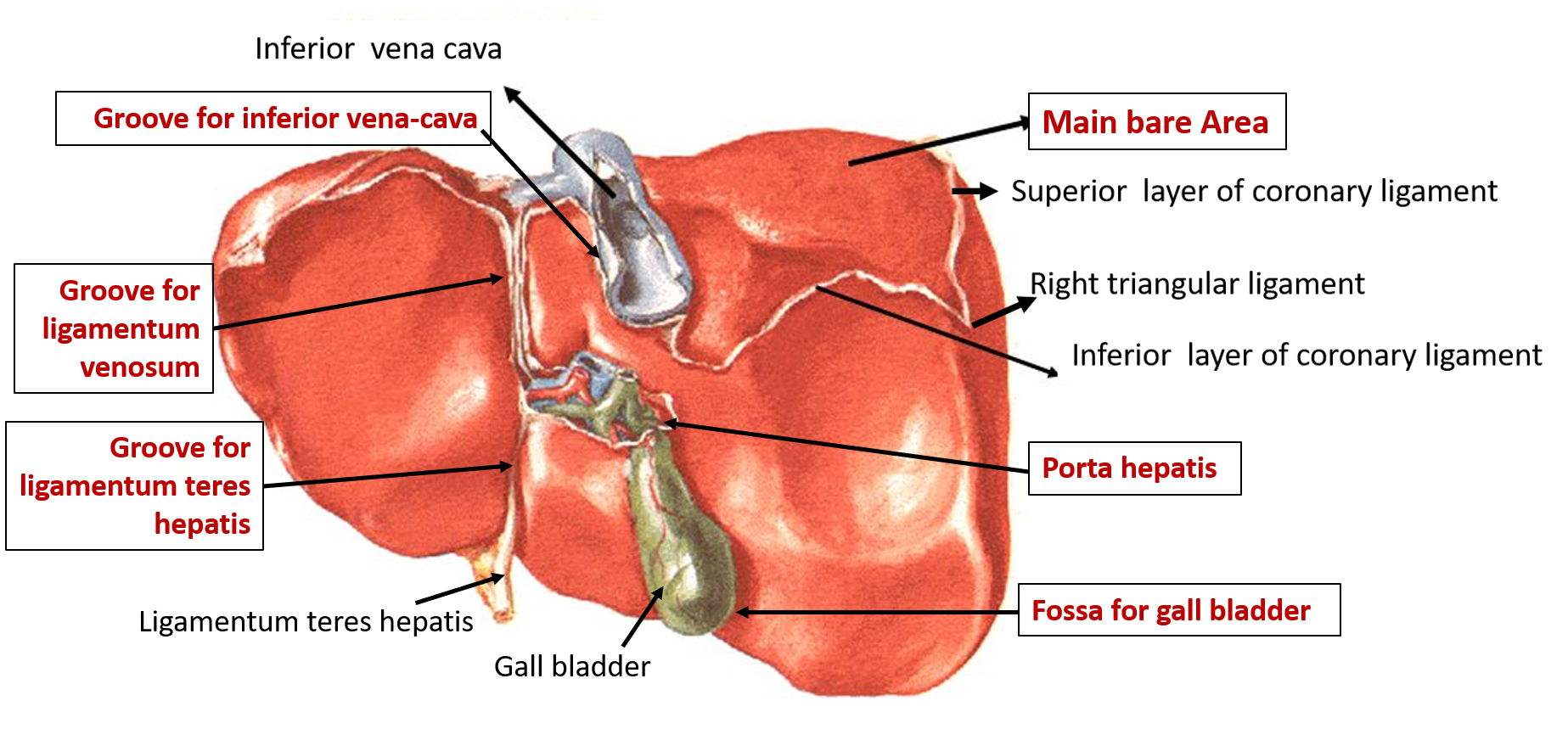
- Main bare area of liver: Is located on the posterior surface. Its boundaries are:
- Base: groove for inferior vena cava
- Apex: right triangular ligament
- Sides: superior and inferior layers of coronary ligaments.
Other bare areas are:
- Groove for inferior vena cava
- Fossa for gall bladder
- Fissures for ligamentum teres hepatis
- Groove/fissure for ligamentum venosum.
- Porta hepatis.
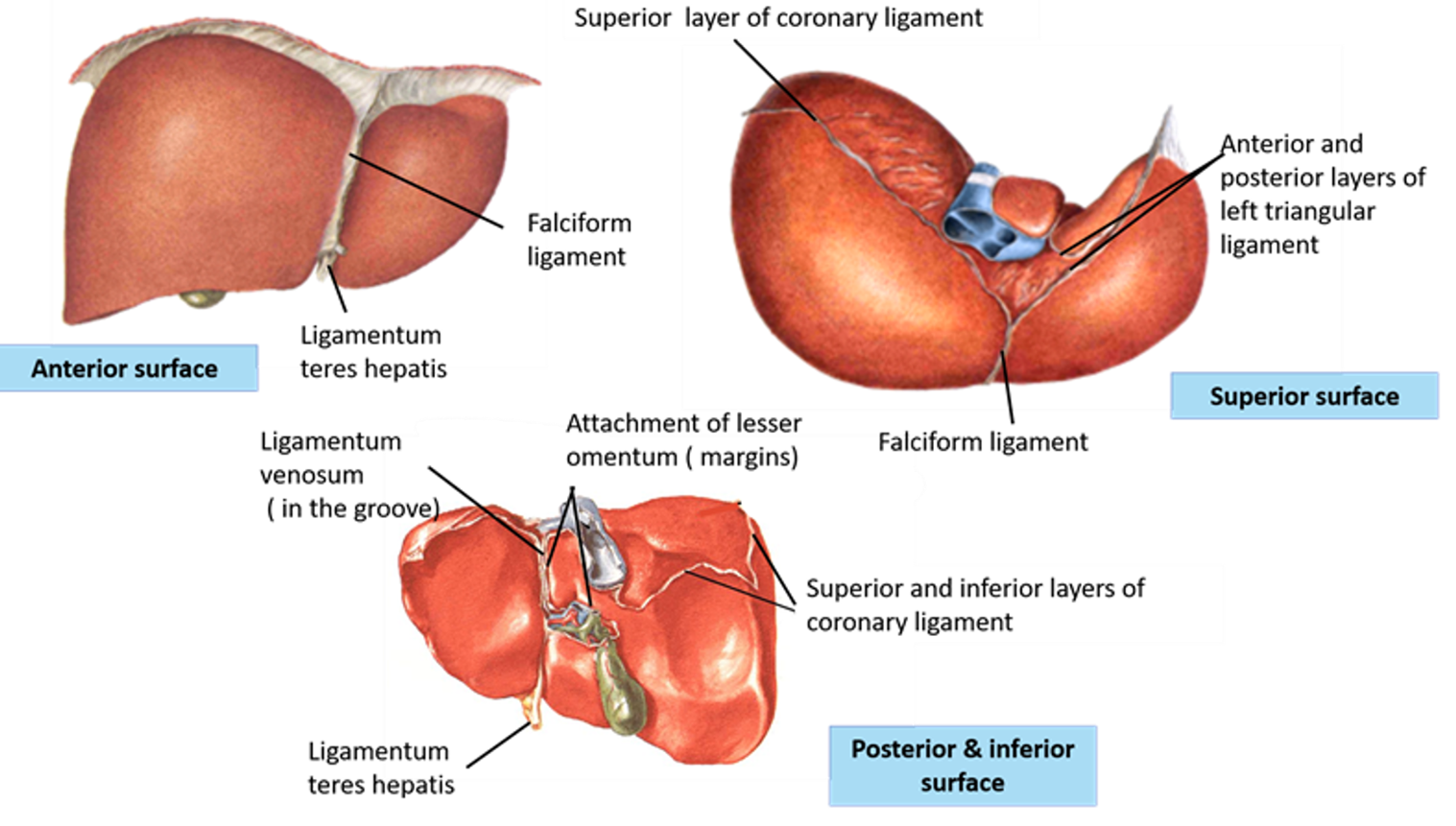
Name the peritoneal folds and ligaments attached to liver.
True ligaments
- Ligamentum teres hepatis (obliterated left umbilical vein), extends from umbilicus to left branch of portal vein.
- Ligamentum venosum (obliterated ductus venosus), extends from left branch of portal vein to inferior vena cava.
False ligaments
- Falciform ligament
- Coronary ligament
- Right and left triangular ligament
- Lesser omentum
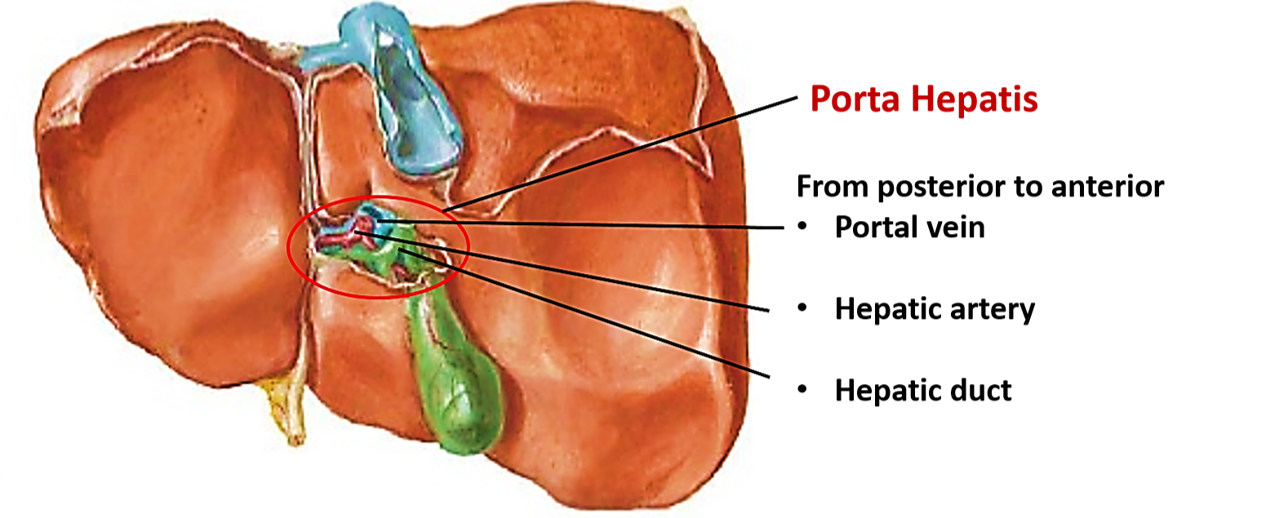
Enumerate the structures passing through the Porta Hepatis.
Structures Passing Through the Porta Hepatis
- Common hepatic duct – anteriorly
- Hepatic artery – in the middle
- Portal vein – posteriorly
- Autonomic nerve fibers (sympathetic from celiac plexus and parasympathetic from vagus)
- Lymph vessels and nodes.
Describe the visceral relations of posterior and inferior surfaces of liver.
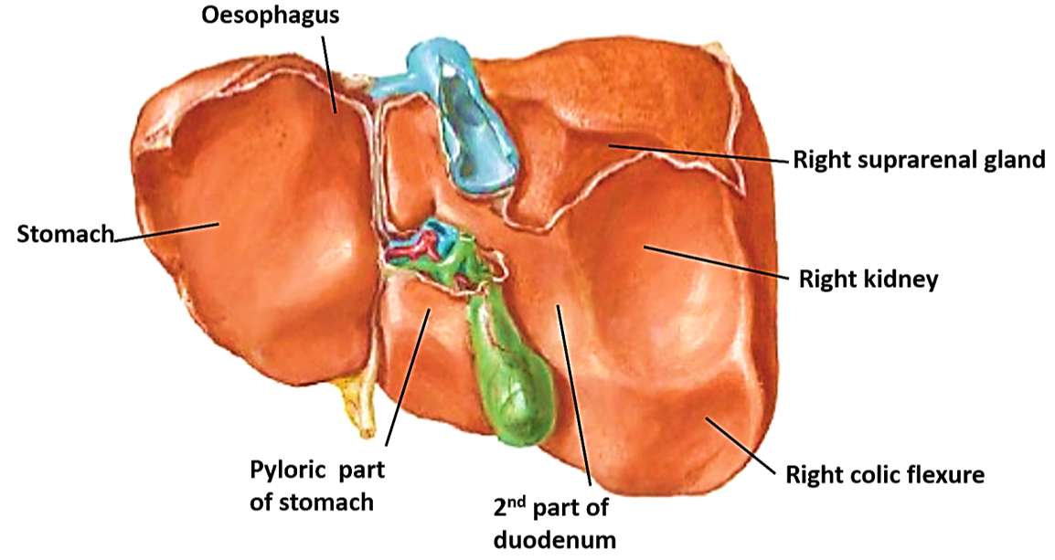
The posterior and inferior surfaces have the following relations:
- On the left of fissure for ligament venosum is related to abdominal part of oesophagus.
- Below this the left lobe is related to the anterosuperior surface stomach (gastric impression).
- Near the left side of the lower end of fissure for ligamentum venosum, the left lobe of liver presents an elevation(tuber omentale/omental tuberosity) which is in touch with the lesser omentum.
- The quadrate lobe is related to the pyloric part of the stomach and the first part of the duodenum.
- The fossa for gallbladder is located to the right of the quadrate lobe.
- The junction of first and 2nd parts of the duodenum is related to the part of the right lobe adjoining gall bladder fossa (duodenal impression).
- Right colic flexure is related to the inferior surface to the right of the gallbladder near the inferior margin (colic impression).
- Right kidney is the inferior surface is related to the inferior surface of right lobe of liver above the colic impression (renal impression).
- Right suprarenal gland is related to lower part of bare area of liver.
Describe the segments of liver.
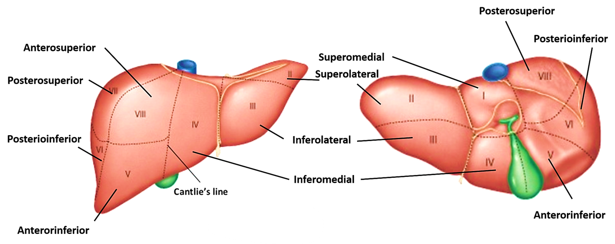
According to Couinaud classification the liver is divided into eight functionally independent segments. In the centre of each segment there is a branch of the portal vein, hepatic artery and bile duct. At the periphery of each segment are the tributaries of hepatic veins The division of the liver into independent units means that segments can be resected without damaging the remaining segments. Segments I-IV are mainly located in the left physiological lobe and Segment V-VII in the right physiological lobe:
- Segment I (Superomedial) is the caudate lobe.
- Segment II (Superolateral) and Segment III (Inferolateral) are located medial to the falciform ligament .
- Segment IV ( Inferomedial) lies lateral to the falciform ligament.
- Segment V (Anteroinferior) is the most medial and inferior.
- Segment VI (Posteroinferior)is located more posteriorly.
- Segment VII (Posterosuperior)is located above segment VI.
- Segment VIII ( Anterosuperior) lies above Segment V.
Applied Aspects
Bare area of the liver
The bare area of the liver is in direct contact with the diaphragm, which separates it from the right pleural cavity. In amoebic hepatitis, the pus may accumulate in this space and create a subphrenic abscess which may burst into the right pleural cavity via the diaphragm.
A potential anastomosis of venous capillaries exists in the region of bare area between the liver and the diaphragm. It becomes functional under certain pathological circumstances (such as portal hypertension).
Biopsy of Liver
In needle biopsy of the liver, the needle is inserted in the midaxillary line through 9th or 10th intercostal space. The needle pierces the thoracic wall, costodiaphragmatic recess of the pleura, diaphragm to reach the right lobe of the liver. Needle inserted above the 8th intercostal space may injure the lung.
High regenerative power of liver
Liver is highly vascular. It has high regenerative power, surgical removal of 2/3rd of liver is compatible with life.
Liver cirrhosis
Liver cells/hepatocytes may under go necrosis following exposure to infection, toxins, alcohol, and poisons. The dead hepatocytes are replaced by fibrous tissue. The resultant hepatic fibrosis is called cirrhosis of the liver. The patient develops jaundice as a result of blockage of bile flow. The increased venous pressure in the portal vein results in portal hypertension which causes engorgement and distension of all its tributaries.

thank for everything
Best website ever
This is the first and best website I have ever seen for anatomy and physiology. One can read all the content specifically. But there is some technical problem with menu please check it.