What is superficial fascia and what are its contents?
Superficial fascia is the subcutaneous layer of loose connective tissue that connects the skin to the underlying deep fascia. It contains:
- Fat
- Superficial blood vessels
- Cutaneous nerves
- Superficial lymphatics
- Mammary gland in females
- Remnants of panniculus carnosus (muscle in superficial fascia) e.g. platysma in neck, muscles of scalp and muscles of facial expression.

What are the functions of Superficial Fascia?
Following are the functions of superficial fascia
- It provides passage to the blood vessels, nerves and lymphatics.
- Acts as cushion and provide insulation (due to presence of fat).
- Allows mobility of skin over underlying structures.
Define Deep Fascia.
Deep fascia is a dense, inelastic fibrous layer that lies deep to superficial fascia and covers the deeper structures such as muscles bone and nerves and blood vessels.
It becomes continuous with the outermost covering layer of underlying structures i.e. periosteum, perimysium, perineurium, and adventitial layer of blood vessels.
What are the Modifications of Deep Fascia?
Modifications of deep fascia are:
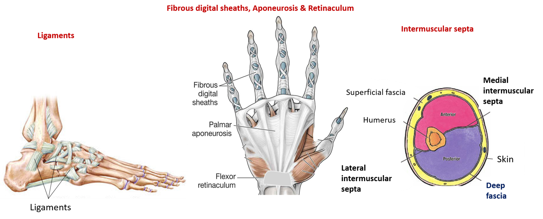
- Intermuscular septa: In limbs the deep fascia sends septa from its deep surface to the bone which separate the muscles into different compartments. They form partition between the groups of muscles.
- Retinaculum: A retinaculum (plural retinacula) is a band of thickened deep fascia that holds tendons in place. They are present where tendons cross the joint. The term retinaculum is derived from the Latin verb retinere (to retain). e.g. Flexor and extensor retinacula are present around the wrist and ankle joint.
- Fibrous flexor sheath: They are thickened deep fascia on the flexor surface of digits in hand and foot. They retain the flexor tendons close to the joints and prevent their bow stringing.
- Ligament: They are the thickening of deep fascia which connect bones at joints. They hold ends of the bones close to each other during movements and thus provide stability to the joints.
- Interosseous membrane: An interosseous membrane is a thick dense fibrous sheet of connective tissue that occupies the space between two bones and connects them. They are present between the radius and ulna bones of forearm and tibia and fibula in the leg.
- Fibrous capsules: Deep fascia forms the external membranous envelope comprising dense, irregular connective tissue, that encapsulates some organs ( such as kidney, liver) and glands (such as parotid, thyroid). these are called fibrous capsule.
- Plantar and palmar aponeurosis : Thickening of deep fascia in sole and palm form plantar and palmar aponeurosis respectively to protect underlying blood vessels and nerves.
Sites where deep fascia is absent : Face and anterior abdominal wall.

Define Bursa and what is Bursitis?
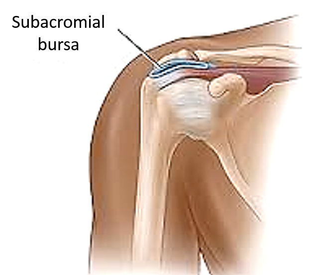
Bursa is a closed serous sac lined by a serous membrane. They are found between the structures that move relative to each other in close apposition. They reduces friction and allow free movement between the apposed surfaces. They are named according to their location e.g. subcutaneous, subtendinous, subcapsular, intertndinous etc.
Infection of bursa is called bursitis.

Adventitious bursae : Bursae that are not normally present but develop over bony surfaces which are subjected to frequent friction and pressure e.g.:
a. Tailor’s ankle: Appears In tailors above the lateral malleolus.
b. Porter’s shoulder : in porters, between upper surface of clavicle and skin.
c. Weaver’s bottom: between gluteus maximus and ischial tuberosity.

Very Nice Notes,Sir do you have pdf or Book or hard copy of your note.