Advertisements
Where is Internal ear Located? What are its Parts?
Location: Internal ear is located in the petrous part of the temporal bone.
Parts of internal ear:
- Internal ear consists of two parts:
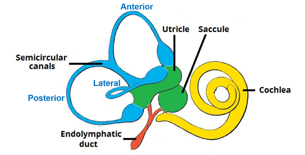
- Outer bony labyrinth. It is filled with a fluid called perilymph
- Inner membranous labyrinth. . It is filled with fluid called endolymph. Membranous labyrinth houses receptors for hearing and equilibrium.
Name the Parts of Bony Labyrinth.
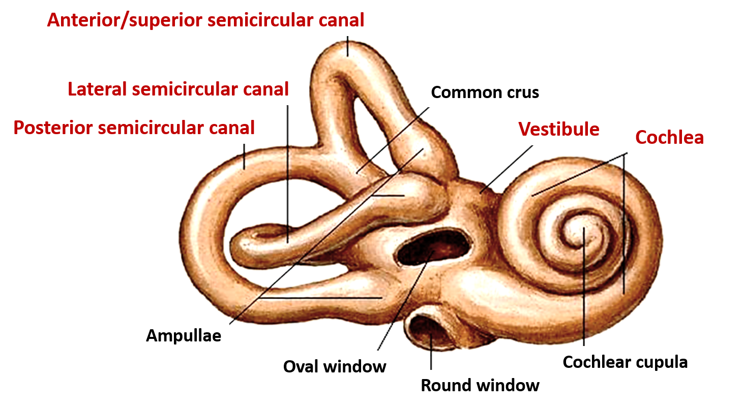
Bony labyrinth consists of three parts:
- Cochlea
- Vestibule
- 3 Semicircular canals
* All contain perilymph, in which the membranous labyrinth is suspended.

Vestibule
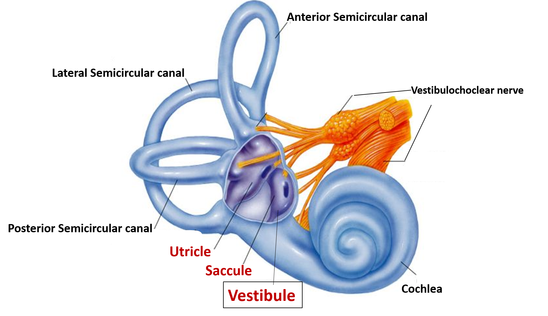
- It is the middle part of the bony labyrinth and contains the utricle and saccule.
- Anteriorly it is continuous with cochlea.
- Posteriorly, it has the openings of semicircular canals.
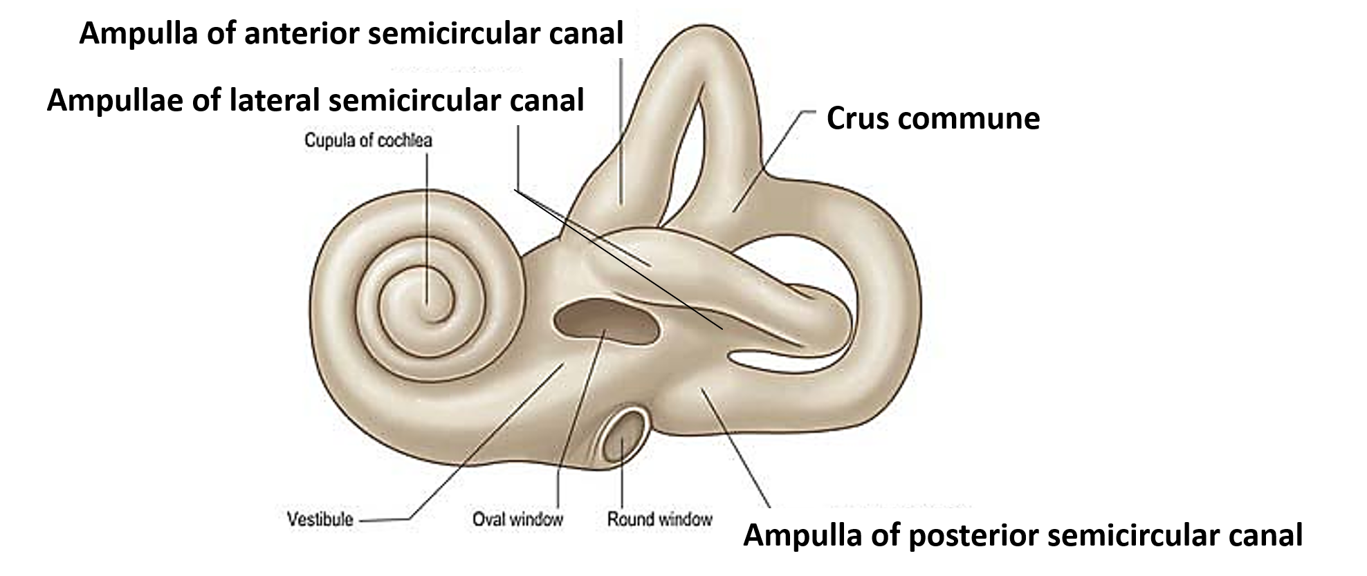
- Its lateral wall has oval window/fenestra vestibuli which is closed by the foot-plate of stapes and annular ligament.
- On its medial wall
- It has a spherical recess anteroinferiorly which lodges saccule and has number of foramina for the lower division of vestibular nerve to reach the saccule.
- It has an elliptical recess posterosuperiorly which lodges utricle and has number of foramina for the upper division of vestibular nerve to reach the utricle.

Cochlea
- It resembles the shell of a snail.
- It is a helical bony tube with 2 and 3/4 turns (coils).
- It has an apex (cupula) directed laterally and a base which is directed towards the internal acoustic meatus.
- Its basal coil forms the promontory on the medial wall of middle ear and it opens posteriorly into the vestibule.
- It contains a central bony pillar called modiolus, from which a spiral ridge projects that partly divides the cochlear canal into two parts:
- Above, scala vestibuli and
- Below, scala tympani
- Two membranes extend from the tip of the spiral lamina to the lateral wall of the chochlea.
- Vestibular membrane stretches from the upper lip of lamina to the outer wall of the cochlea.
- Basilar membrane stretches from the lower lip of the lamina to the outer wall of the cochlea.
- The triangular area enclosed by the vestibular and basilar membranes and the outer wall of the cochlear canal enclose the cochlear duct (scala media).
- Scala vestibuli and tympani communicate with each other at the apex of cochlea via a small opening called helicotrema. Scala tympani is closed by bony lamina (at the round window), while the scala vestibuli opens into the vestibule.

Semicircular canals
- There are three semicircular canals viz. anterior (superior), lateral and posterior.
- Each semicircular canal is 15-20 mm long.
- The three canals are at right angle to each other.
- Each canal is dilated at both ends to form ampullae.
- They open into vestibule via 5 openings (posterior end of anterior and the upper end of posterior semicircular canals join to form crus communae which opens via single opening).

Describe the Membranous Labyrinth.
Membranous labyrinth contains endolymph and has following parts:
- Utricle and saccule are present in the vestibule and they contain sense organs called maculae which detect linear acceleration of the head.
- 3 semicircular ducts are present in the 3 semicircular canals, their dilated ends are called ampullae have crest which contain receptors to detect rotational acceleration .
- Cochlear duct is present within the cochlea which contains organ of Corti, which is responsible for sense of hearing.

Describe Briefly the Blood and Nerve Supply of Internal Ear.
Arterial Supply: Following arteries supply internal ear:
- Labrynthine artery (branch of basilar artery).
- Stylomastoid branch of occipital artery.
Organ of corti has no blood vessels.
Venous Drainage: Veins of internal ear drain into the superior petrosal sinus.
Nerve Supply:
- Urticle, saccule and semicircular canals receive fibers from vestibular nerve.
- Cochlear duct ( organ of Corti) receives fibers from cochlear nerve.
