Where is tongue loacted? What are its Functions?
Tongue is a mobile muscular organ, essentially made up of skeletal muscle covered by mucous membrane.
Location: It is partly located in the oral cavity and partly in oropharynx.
Functions of tongue : The functions of tongue are:
- taste
- speech(articulation)
- mastication
- swallowing.
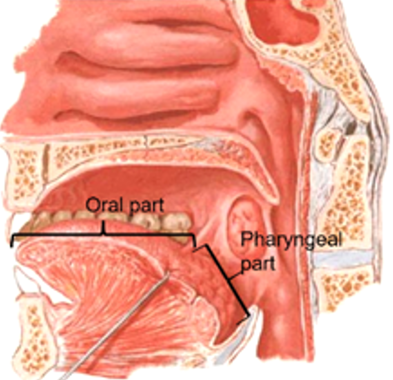
What are the Parts of Tongue?
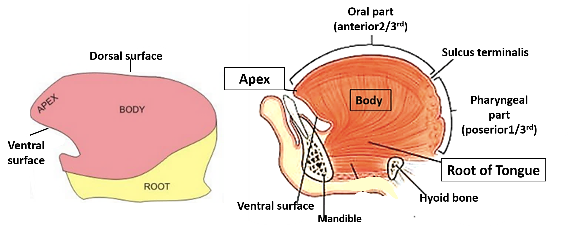
Tongue consists of following parts:
- Tip/Apex: It is directed forward and is in contact with incisors when the mouth is closed.
- Root: Lies in the floor of mouth and is attached to mandible and hyoid bone by muscles.
- Body: is the part between the tip and root. It has:
- two surfaces – ventral (inferior) and dorsal (superior) and
- two lateral borders/margins.
Describe the Features of Ventral Surface of Tongue.
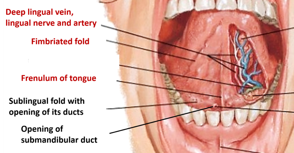
Ventral surface of tongue is covered by mucous membrane which is continuous with that of floor of mouth. It shows following features:
- Frenulum linguae : midline mucosal fold which connects the ventral surface to floor of mouth.
- Deep lingual vein: located on either side of frenulum linguae dep to mucosa.
- Plica fimbriata: fimbriated mucosal fold lateral to deep lingual veins.
Describe the Features of Dorsal Surface of Tongue.
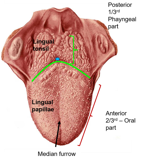
Dorsal surface of tongue presents a V-shaped sulcus (sulcus terminalis). At the apex of sulcus terminalis there is a depression called foramen caecum. Foramen caecum indicates the site of origin of thyroid diverticulum (thyroglossal duct).
Sulcus terminalis divides the dorsal surface into two parts:
- Anterior 2/3rd (oral part): This part presents a median furrow and the surface has a rough appearance. The mucous membrane is characterized by presence of surface projections called lingual papillae.
- Posterior 1/3rd (pharyngeal part): It forms the anterior wall of oropharynx. It is connected to palate by palatoglossal arches (containing palatoglossus muscle. A median and two lateral glossoepiglottic folds connect it to epiglottis. The mucosa of this part has no papillae. It has large number of lymphoid follicles (lingual tonsil) deep to mucosa.
<
What are the Types of Lingual Papillae?
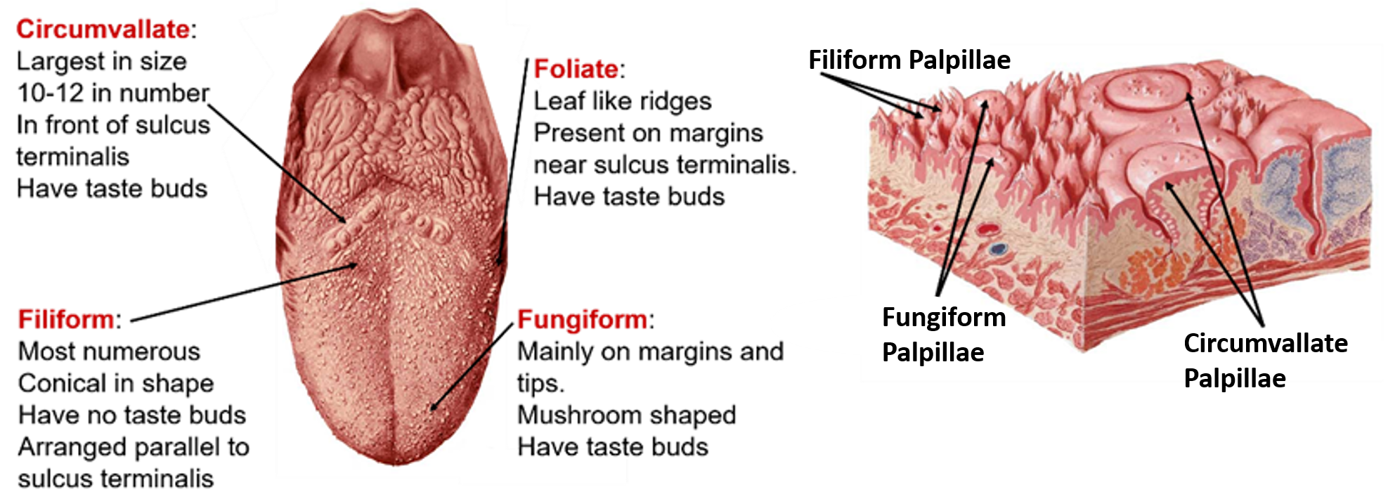
Mucus membrane of anterior 2/3rd of tongue has four types of lingual papillae. Lingual papillae are projections of lamina propria of mucosa covered by stratified squamous epithelium. The four types of papillae are:
- Filiform papillae:
- Are conical in shape.
- Epithelium covering them is keratinized.
- Are arranged in rows parallel to sulcus terminalis.
- Are devoid of taste buds.
- Fungiform papillae:
- Are rounded at top and narrow at base.
- Are mostly present at tip and margins of tongue.
- Have taste buds.
- Circumvallate papillae:
- Are largest in size -2-3 mm (10-12 in number).
- Are located in front of the sulcus terminalis.
- Their shape is like truncated cone and are surrounded by a circular sulcus.
- Have numerous taste buds on the sides.
- Foliate papillae:
- Are leaf like transverse ridges present on the lateral margin of tongue in front of sulcus terminalis.
- They bear taste buds.
Describe Origin, Insertion, Action and Nerve Supply of Tongue.
The muscle mass of tongue is divided into two halves. Each half of the tongue contains four intrinsic and four extrinsic muscles.
Intrinsic muscles are limited to the upper part of the tongue and are responsible for changing the shape of the tongue. They are:
- Superior longitudinal
- Inferior longitudinal
- Transverse
- Verticalis
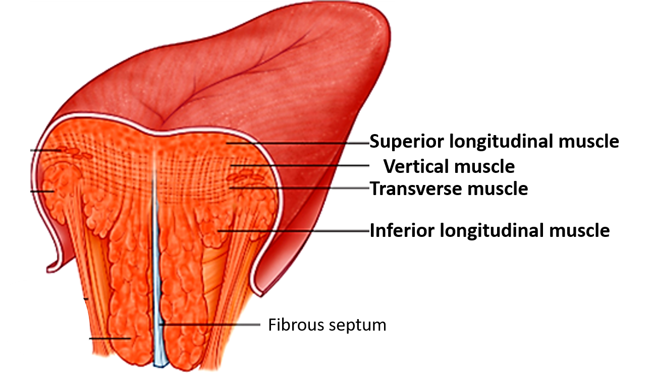
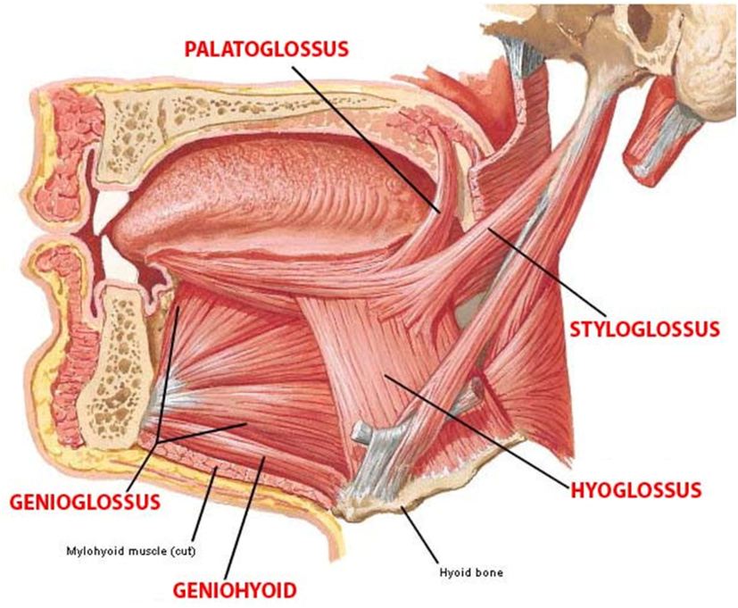
Extrinsic muscles are responsible for moving the tongue and they attach the tongue to:
- Mandible anteriorly (Genioglossus)
- Palate superiorly (Palatoglossus)
- Styloid process posteriorly (Styloglossus)
- Hyoid bone inferiorly (Hyoglossus)
| Muscle | Action | Nerve Supply | |
|---|---|---|---|
| Intrinsic Muscles | Superior longitudinal | Shorten the tongue and make the dorsal surface concave | |
| Inferior longitudinal | Shorten the tongue and make the dorsal surface convex | ||
| Transverse | Make the tongue narrow and elongated | All intrinsic muscles are supplied by hypoglossal (XII cranial) nerve | |
| Vertical | Make the tongue flat and broad | ||
| Extrinsic Muscles | Genioglossus | Protrude the tongue (when both the muscles contract). | All the muscles are supplied by hypoglossal nerve except palatoglossus which is supplied by vago-accessory complex via pharyngeal plexus. |
| Hyoglossus | Depress the side of the tongue | ||
| Styloglossus | Pulls the tongue upwards and backwards | ||
| Palatoglossus | The two muscles together approximate the palatoglossal arches thereby closing the oropharyngeal isthmus |
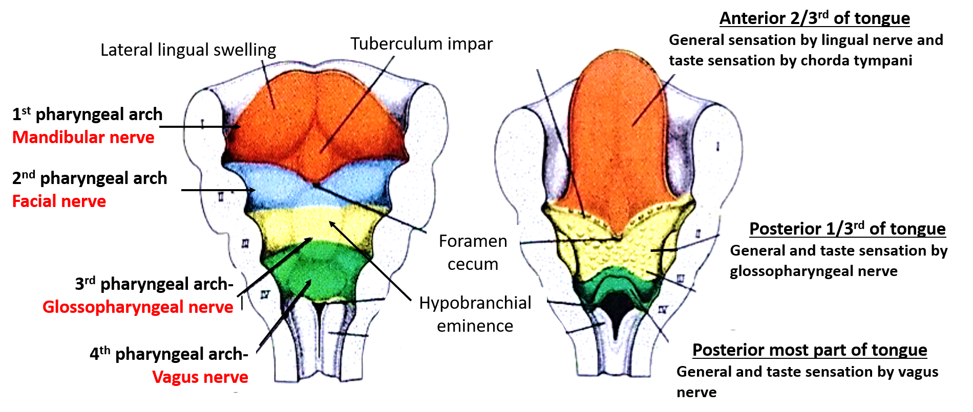
Describe the Sensory Nerve of Tongue and Correlate it with It’s Development.
| Part of the tongue | Nerve carrying general sensation | Nerve carrying taste sensation | Correlation with Development |
|---|---|---|---|
| Anterior 2/3rd of the tongue (except circumvallate papillae) | Lingual nerve (branch of mandibular nerve) | Chorda tympani (branch of facial nerve) | Mucosa of anterior 2/3rd of tongue develops from 1 st pharyngeal arch, therefore, general sensory innervations is by mandibular nerve (nerve of 1 st arch). |
| The pre-trematic nerve of first arch is facial nerve, therefore taste sensation is carried by chorda tympani, branch of facial nerve. | |||
| Posterior 1/3rd of the tongue (including circumvallate papillae) | Glossopharyngeal nerve | Glossopharyngeal nerve | Mucosa of the posterior 1/3 rd of tongue develops from 3rd pharyngeal arch, therefore supplied by nerve of third arc i.e. glossopharyngeal nerve. |
| Posterior most part of tongue including vallecula. | Internal laryngeal nerve (branch of vagus) | Internal laryngeal nerve (branch of vagus) | Mucosa of the vallecula of tongue develops from 4th pharyngeal arch, therefore supplied by nerve of 4 th pharyngeal arch i.e. vagus nerve |
Motor supply of tongue: Most of the muscles of tongue develop from occipital myotomes and is supplied by hypoglossal nerve
Describe Blood Supply of Tongue.
Arterial supply of tongue
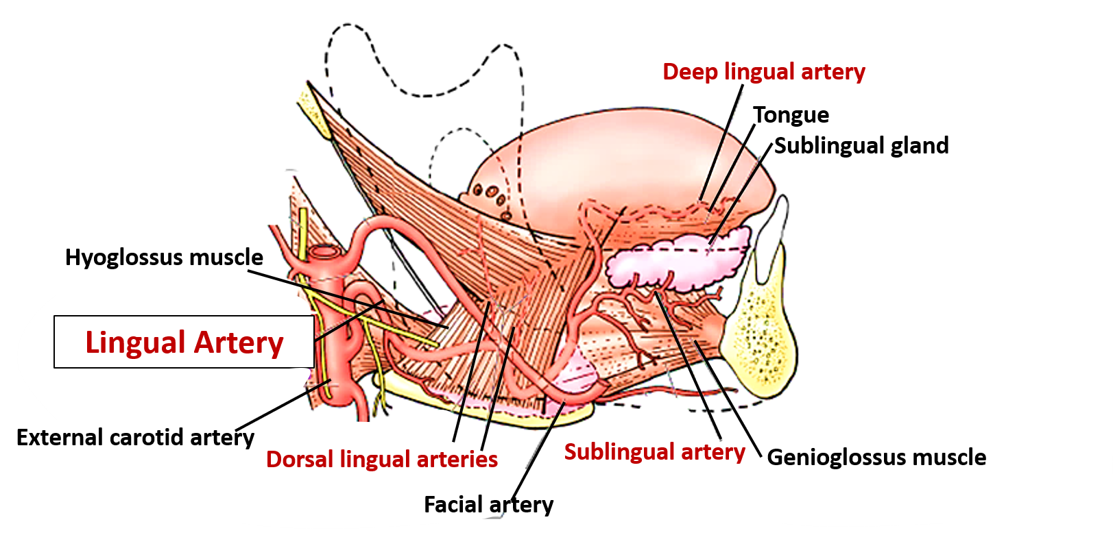
- Lingual artery: a branch of external carotid artery(ECA) is the chief artery that supplies tongue. It arises from the anterior aspect of ECA opposite the tip of the greater cornu of hyoid bone. It is divided into three parts by hyoglossus muscle:
- 1st part: Lies in the carotid triangle. Forms a loop above the greater cornu.
- Branch: suprahyoid artery.
- 2nd part: passes deep to hyoglossus muscle.
- Branches: dorsal lingual arteries (usually two) supply posterior part of the tongue and palatine tonsil.
- 3rd part: also called deep lingual artery or arteria profunda linguae runs upwards along the anterior border of hyoglossus and then forward on the under surface of tongue.
- Branches: sublingual artery to sublingual gland and branches to anterior part of tongue.
Venous drainage of tongue
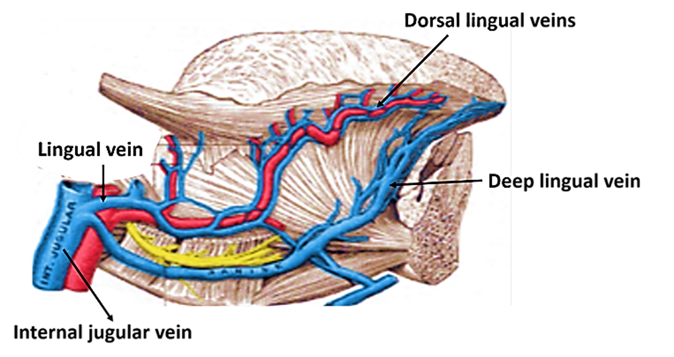
- Dorsal lingual veins: drain the dorsum and sides of the tongue run and join the venae commitantes accompanying lingual artery to form lingual vein .
- Deep lingual veins of tongue drain the tip and form the venae commitantes accompanying the hypoglossal nerve. These veins unite at the posterior border of hyoglossus which may drain into lingual vein or into internal jugular vein.
Describe the Lymphatic Drainage of Tongue.
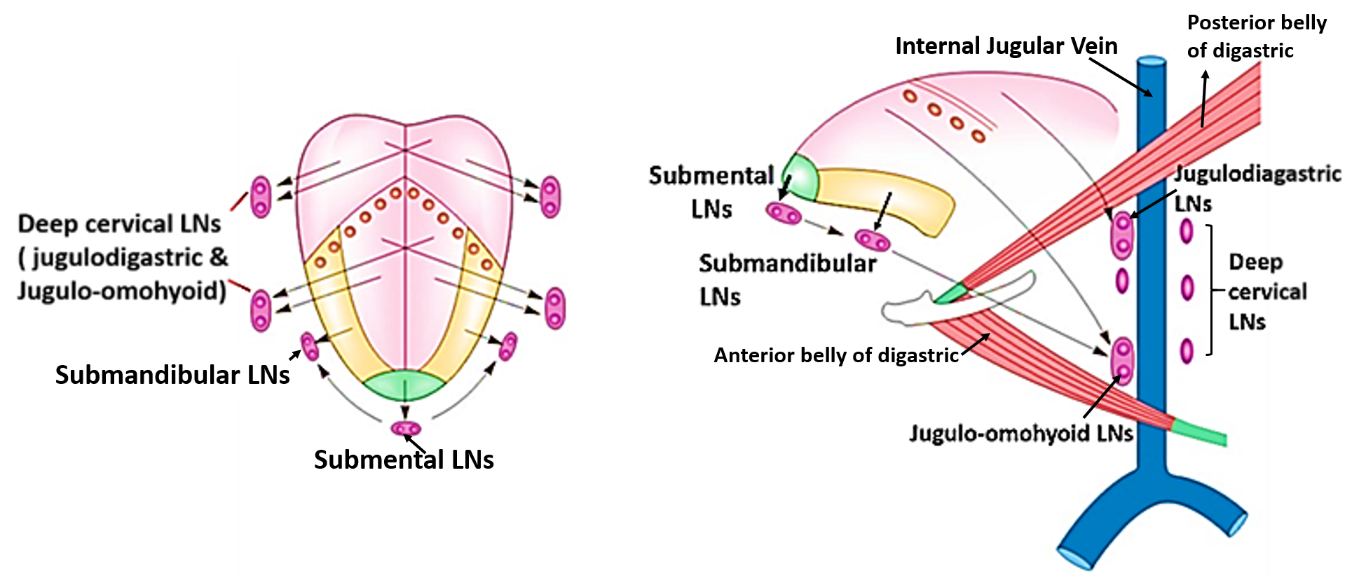
Lymphatic drainage of tongue: Lymphatic vessels draining tongue are divided into the following groups:
- Apical vessels: drain the tip of the tongue into submental lymph nodes which may drain into submandibular or jugulo-omohyoid lymph nodes.
- Marginal vessels: drain the lateral (marginal) area of anterior 2/3rd of tongue unilaterally into the submandibular lymph nodes which then drain into lower group (jugulo-omohyoid) of deep cervical lymph nodes..
- Central vessels: drain the central part of anterior 2/3rd of tongue bilaterally into deep cervical lymph nodes including jugulo-omohyoid lymph nodes.
- Basal vessels: drain posterior 1/3rd of the tongue bilaterally into the jugulo-diagastric and retropharyngeal lymph nodes.
Main lymph nodes draining tongue are Jugulo-omohyoid lymph nodes and those for palatine tonsil are Jugulo-digastric.
Applied Aspect
Malignant tumors of the posterior 1/3rd have poor prognosis
There is rich anastomosis across the midline between the lymphatics of posterior 1/3rd of tongue. Therefore, tumors cells of one side readily metastasize to contralateral side.Tumors located more than ½ inch from midline do not metastasize to the opposite side till late as there is little cross communication between the lymphatics of the two sides.
Genioglossus is known as the ‘safety muscle of the tongue’
Genioglossus is known as the ‘safety muscle of the tongue’ because it protrudes the tongue and prevents it from falling back and thereby blocking airway in an unconscious patient.
