What are the characteristic features of skin and fascia of Face?
Characteristic features of skin of face are:
- It is highly vascular and elastic.
- It contains large number of sweat and sebaceous glands (is common site for acne).
- It is lax except on nose where it is attached to the underlying cartilages (laxity of the skin facilitates rapid spread of edema).
- It provides insertion to the muscles of facial expression.
Superficial Fascia: It contains muscles of facial expression, vessels and nerves and varying amount of fat. The fat is absent in the eyelids , however it is abundant in cheeks (especially in children) and is called buccal pad of fat.
♦ Deep fascia : It is absent in face except over the parotid gland and masseter muscle (parotido-masseteric fascia).The absence of deep fascia in the face allows facial expressions to be seen.
What are the special features of muscles of facial expression?
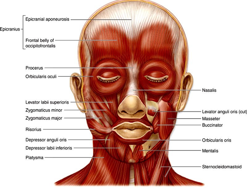
- The muscles of facial expression are present in the superficial fascia of the face.
- Most of them take origin from bones of facial skeleton and are inserted into the skin.
- Morphologically, they represent the subcutaneous muscle (panniculus carnosus) present in some animals.
- Embryologically, they arise from the mesoderm of second pharyngeal/branchial arch and are therefore supplied by facial nerve, the nerve of second arch.
- Functionally, they are are arranged around three orifices palpebral fissure, nostrils and mouth and are responsible for their closure (sphincters) of opening (dilators).
- They are also responsible for facial expressions.
Write in a tabulated form the origin, insertion and action of Muscles of facial Expression

Muscles around palpebral fissure
| Muscle | Origin | Insertion | Action |
|---|---|---|---|
| Orbicularis oculi (Orbital part) | Medial palpebral ligament, frontal process of maxilla and adjoining part of the frontal bone | Muscle fibers form elliptical loops around the orbital margin and return to the point of origin | Closes eyelids tightly (as in winking, protection from light) |
| Orbicularis oculi (Palpaberal part) | Medial palpebral ligament | Muscle fibers pass in the upper and lower eyelids and are inserted into the lateral palpebral ligament | Closes eyelids gently (as in blinking or sleep) |
| Orbicularis oculi (Lacrimal part) | Posterior lacrimal crest and lacrimal fascia | Muscle fibers pass through both eyelids to be attached to the lateral palpebral raphe | Dilates lacrimal sac and helps and aids in drainage of lacrimal fluid |
| Corrugator supercilii | Medial end of the superciliary arch | Fibers run laterally and upwards to be inserted into the skin of the eyebrow | Produce vertical lines in the forehead (as in irritation) |
Although Levator palpabrae superioris is not a muscle of the face however, it acts as an antagonist to the sphincteric activity of orbicularis oculi. It elevates the upper eyelid. It is supplied by Oculomotor nerve.
Muscles around nose
| Muscle | Origin | Insertion | Action |
|---|---|---|---|
| Procerus | Nasal bone | Skin of the medial part of eyebrow | Produces transverse wrinkles over the bridge of the nose |
| Compressor naris | Maxilla near the nasal notch | Passes medially to form an aponeurosis via which it becomes continuous with its counterpart on the opposite side | Compresses the nasal aperture |
| Dilator naris | Maxilla from the margin of the nasal notch | Skin of the ala of the nose | Draws the ala downwards and laterally thus assists in widening the nasal aperture (as in deep inspiration and anger) |
| Depressor septi | Incisive fossa of the maxilla | Lower mobile part of the nasal septum | Widens the nasal aperture. |
Muscles around Mouth
Nine Muscles converge around the mouth. These are as follows:
- Levator labii superioris alaeque nasi.
- Levator labii superioris.
- Levator anguli oris.
- Zygomaticus minor.
- Zygomaticus major.
- Depressor labii inferioris.
- Depressor anguli oris.
- Risorius.
- Buccinator
| Location | Muscle | Origin | Insertion | Action |
|---|---|---|---|---|
| Around mouth | Orbicularis oris | Extrinsic part from other muscles around the mouth and intrinsic part from incisive fossae | Encompasses the oral orifice | Closes the mouth |
| Muscles attached to the upper lip | Labial part of levator labii superioris alequae nasi | Frontal process of the maxilla | Upper lip | Elevates upper lip |
| Levator labi superioris | Maxilla just above the infraorbital foramen | Upper lip | Elevates and everts upper lip | |
| Zygomaticus minor | Zygomatic bone | Upper lip | Elevates upper lip | |
| Muscles converging at the angle of mouth | Levator anguli oris | Maxilla below the infraorbital foramen | Angle of the mouth | Raises the angle of mouth |
| Zygomaticus major | Zygomatic bone | Angle of the mouth | Pulls the angle of mouth upwards and laterally | |
| Depressor anguli oris | Oblique line of the mandible | Angle of the mouth | Draws the angle of mouth downwards and laterally | |
| Risorius | Parotid fascia as continuation of posterior fibres of platysma | Angle of the mouth | Pulls the angle of mouth laterally | |
| Muscle attached to the lower lip | Depressor labii inferioris | Oblique line of the mandible | Lower lip | Draws the lower lip downwards |
| Platysma | Fascia over the upper parts of pectoralis major and deltoid muscles | Base of the mandible and lower lip | Draws down the lower lip and angle of mouth | |
| Mentalis | Incisive fossa of the mandible | Skin of chin | Protrudes or everts the lower lip (although it is not attached to lower lip) |
Name the muscles responsible for various Emotional Expressions.
| Facial expression | Muscle |
|---|---|
| Frowning | Corugator supercilii and procerus |
| Surprise | Frontalis |
| Laughing | Zygomaticus major |
| Grinning | Risorius |
| Grief | Depressor anguli oris |
| Anger | Dilator naris and depressor septi |
| Doubt | Mentalis and Risorius |
Write the origin, insertion, action and nerve supply of Buccinator Muscle.
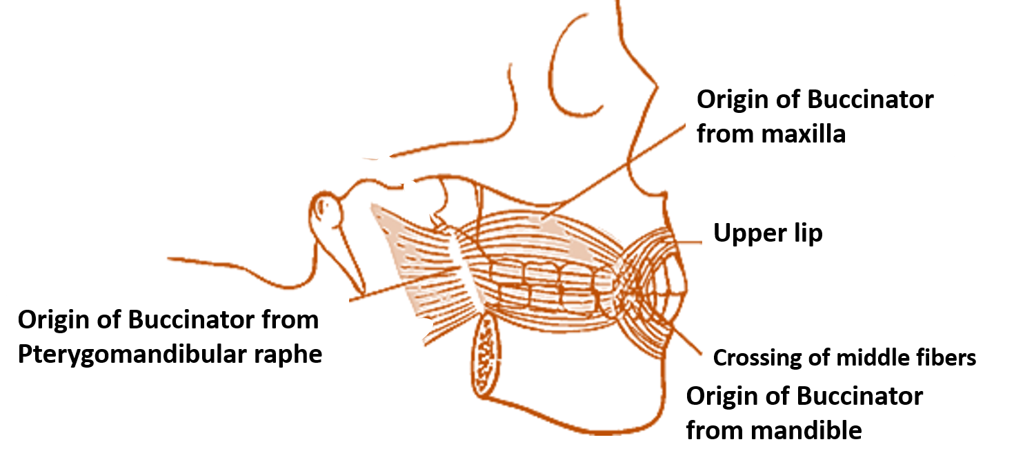
Buccinator (Trumpeter’s muscle) is a quadrilateral shaped muscle of cheek.
| Origin | Insertion | Action | Nerve Supply |
|---|---|---|---|
| Upper fibers from the outer surface of maxilla opposite to the molar teeth | Upper lip | Compresses the cheek against teeth and gums. This prevents accumulation of food in the vestibule of mouth. Helps in whistling, blowing. | Buccal branch of facial nerve |
| Middle fibers from the Pterygomandibular raphe | Fibers decussate at the angle of mouth and upper fibers pass to lower lip , lower fibers to upper lip. | ||
| Lower fibers from the outer surface of mandible opposite to the molar teeth | Lower lip |

Structures piercing buccinator muscle:
- Parotid duct
- Buccal branch of mandibular nerve.
Applied Aspects
Facial plastic surgery is mostly successful
As skin of the face has very rich blood supply, it is uncommon in plastic surgery for skin flaps to necrose.
Edema spreads fast on face
The laxity of skin over most of the parts of face allows fast spread of edema in the region of the face.
Face is common site for acne
It is due to the presence of large number of sebaceous glands in this region.
What is Ectropion and Epiphora?
Paralysis of orbicularis oculi leads to drooping of the lower eyelid called ectropion, which causes spilling of tear on the cheek (Epiphora).
What happens in case of paralysis of buccinator muscle?
Paralysis of buccinator muscle (in facial palsy – injury to facial nerve), food tends to accumulate in the vestibule of mouth and the person is unable to blow and whistle.
For Bell’s Palsy (Click here)
