Describe the location, extent and gross features of kidney.
Location: kidneys are located retroperitoneally on the posterior abdominal wall on either side of vertebral column.
Extent: It extends from T12 to L3 vertebra (right kidney is slightly lower than the left).
* The transpyloric plane (lower border of L1 vertebra) passes through the upper part of the hilum of right kidney and lower part of the hilum of left kidney.
Gross features:
- Shape: Bean shaped.
- Colour: Reddish brown
- Dimensions: Weight- 130-170gm, Length – 11cm., Breadth – 6cm., Thickness – 3cm.
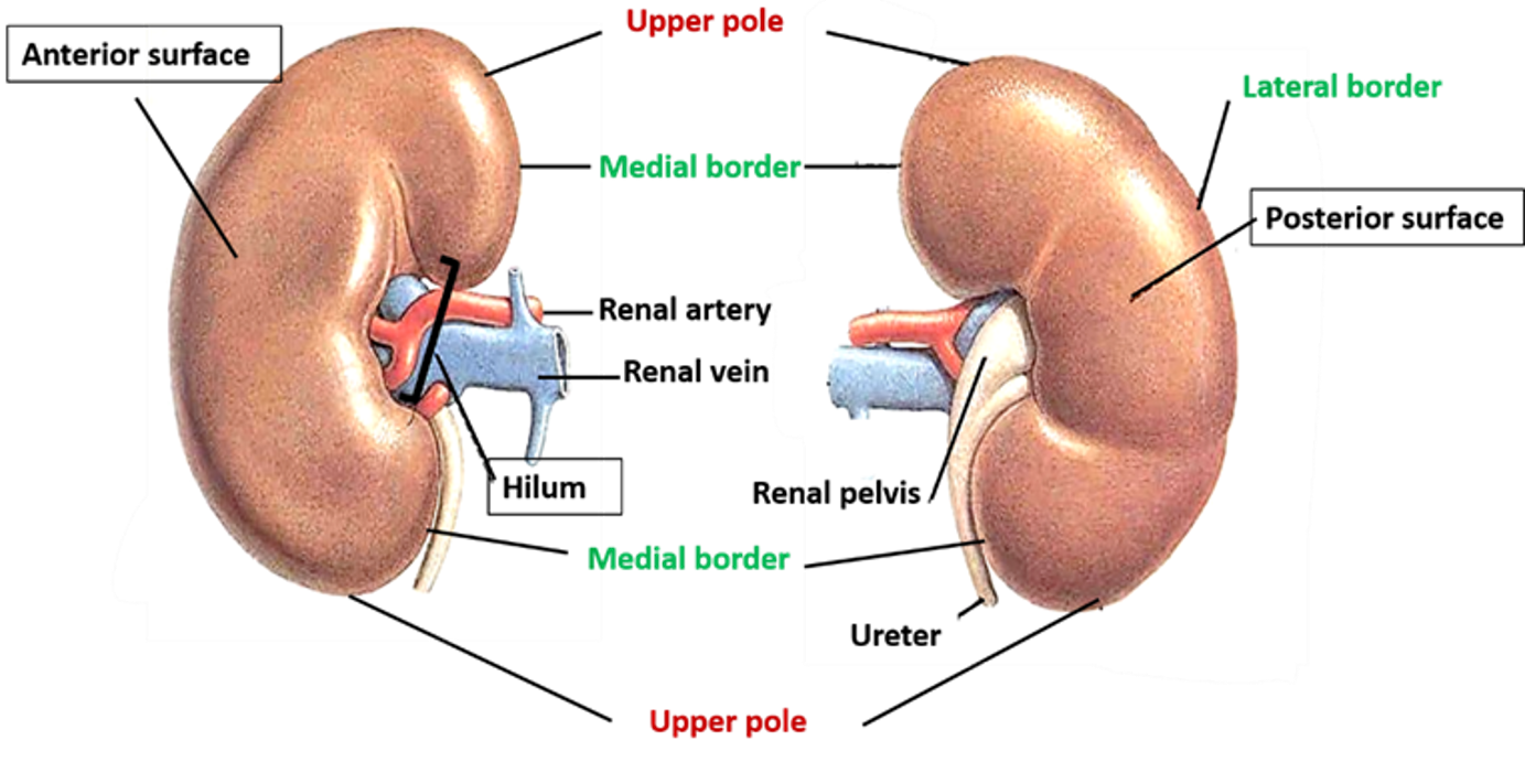
Each kidney has:
- Two surfaces:
- Anterior: is convex and faces anterolaterally.
- Posterior: is flat and faces posteromedially.
- Two poles:
- Upper pole: is nearer to the median plane than the inferior pole.
- Lower pole: is more laterally placed as compared to upper pole and is about 1 inch above the iliac crest.
- Two borders:
- Lateral border: is convex.
- Medial border: It presents a vertical cleft in the middle called hilum.
- Hilum: Is present along the central part of medial border. The structures passing through it from before backwards are:
- Renal vein
- Renal artery
- Ureter (actually renal pelvis)
Describe the coverings of kidney.
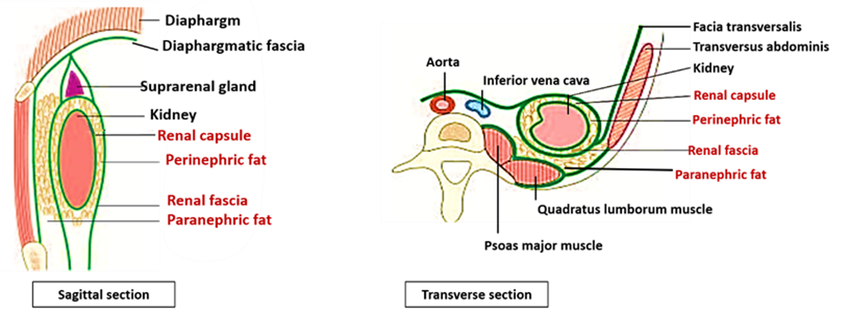
From within outwards the coverings are:
- Fibrous capsule: Covers the entire organ, lines the renal sinus and covers the walls of major and minor calyces and pelvis of ureter.
- Perinephric fat: Is abundant along the borders of the kidney and extends through the hilum into the renal sinus.
- Renal fascia/Fascia of Gerota: Is a fibroareolar sheath which surrounds the kidney and perirenal fat. It consists of two layers –anterior and posterior.
- Above: the two layers fuse at the upper pole and split again to enclose the suprarenal gland & then blend with the subdiaphragmatic fascia.
- Laterally: Both the layers fuse and become continuous with the fascia transversalis.
- Medially: Anterior layer merges with the connective tissue surrounding aorta and inferior vena cava. Posterior layer is continuous with the psoas fascia. Along the medial border of the kidney the two layers are connected to each other by a connective tissue septum.
- Below: The two layers do not fuse and extend downwards along the ureter. Anterior layer merges with extraperitoneal tissue of iliac fossa and the posterior layer with fascia iliaca.
- Paranephric fat: It is a layer of fat present outside the renal fascia. The fat in this layer is more abundant posteriorly between the thoracolumbar fascia and renal fascia.
Describe the macroscopic structure of kidney.
Naked eye examination of a coronal section of kidney shows:
- Renal cortex: It is the outer part of the kidney, located just below the capsule. Bowman’s Capsules and the glomeruli, proximal and distal convoluted tubules are located in this part. It is divided into two parts:
- Outer cortical lobules- over the renal pyramids
- Renal columns extending between pyramids of renal medulla.
- Renal medulla: It is the inner part of the kidney. It has 10-12 dark coloured pyramids, The base of each pyramid is towards the cortex and the apex (renal papilla) opens in minor calyx. It contains loops of Henle, collecting tubules and collecting ducts. Extension of medulla into the cortex forms medullary rays (containing collecting tubules).
Each medullary pyramid and the associated cortical tissue at its base and sides (one half of each adjacent renal column) constitutes a lobe of the kidney.
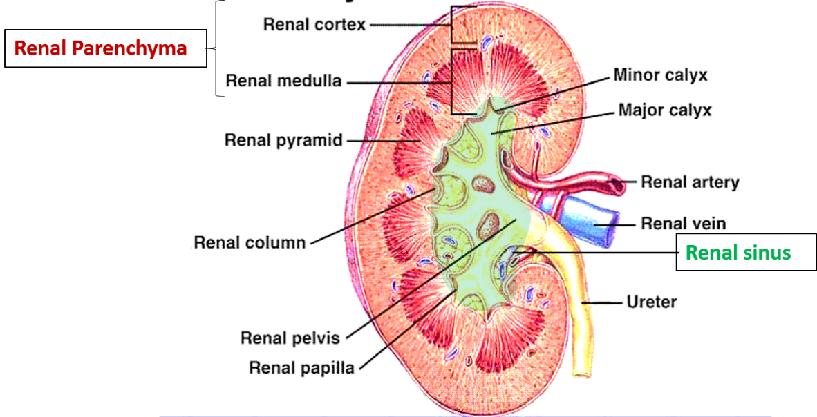
Renal sinus: It is a space between the renal parenchyma and hilum. It contains:
- Minor calyces
- Major calyces
- Pelvis of kidney (which at the lower pole of kidney continues as ureter)
- Blood vessels and nerves
- Perinephric fat
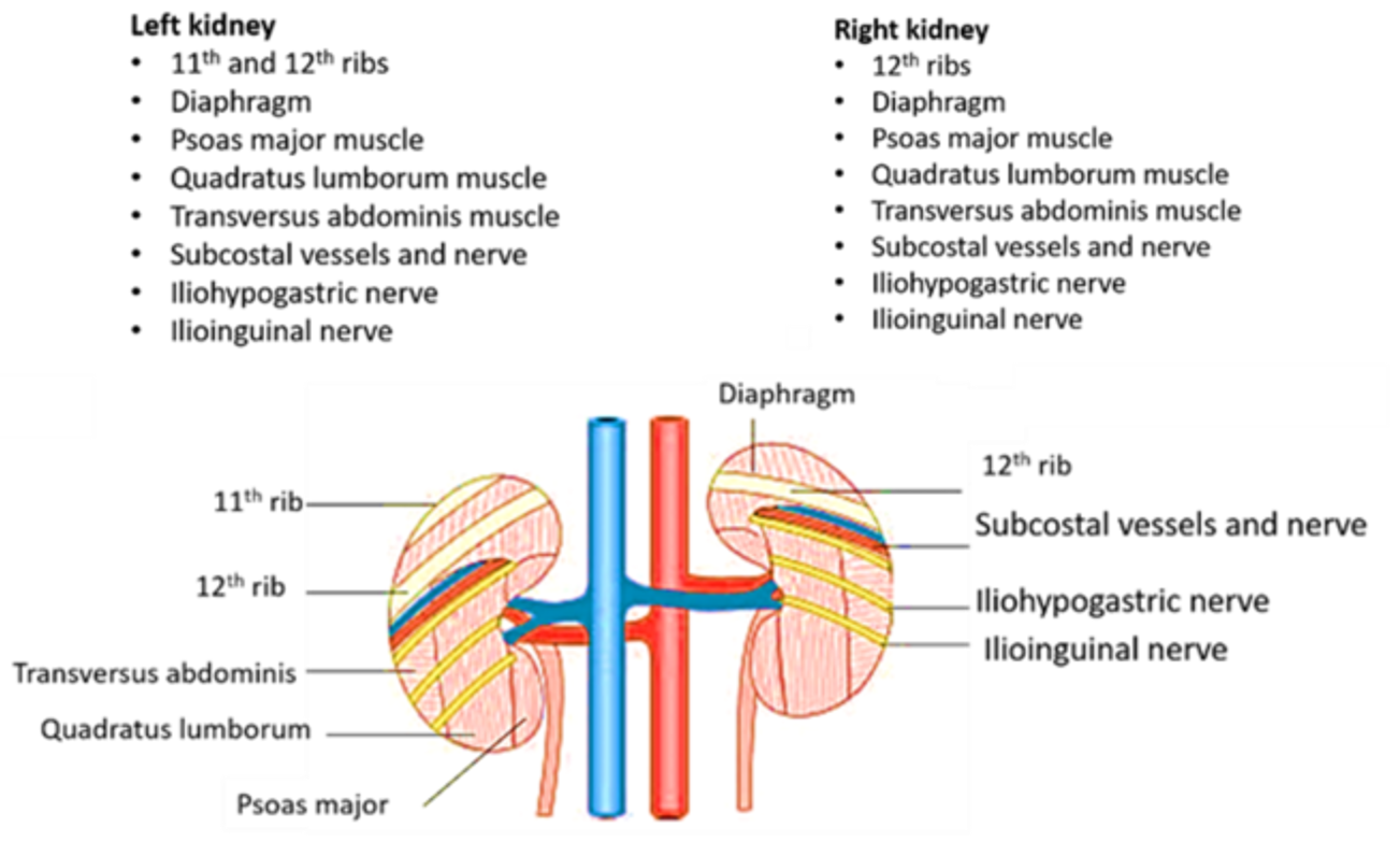
What are the relations of kidney?
Anterior relations of right and left kidneys
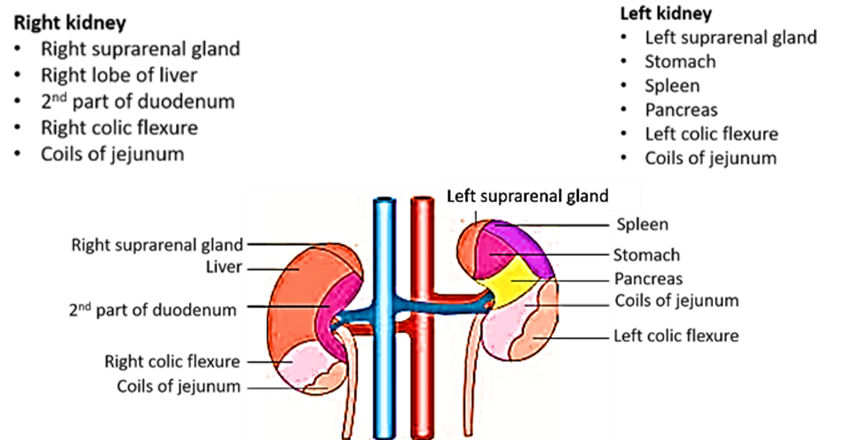
Posterior relations of right and left kidneys
Describe the blood supply of kidney.
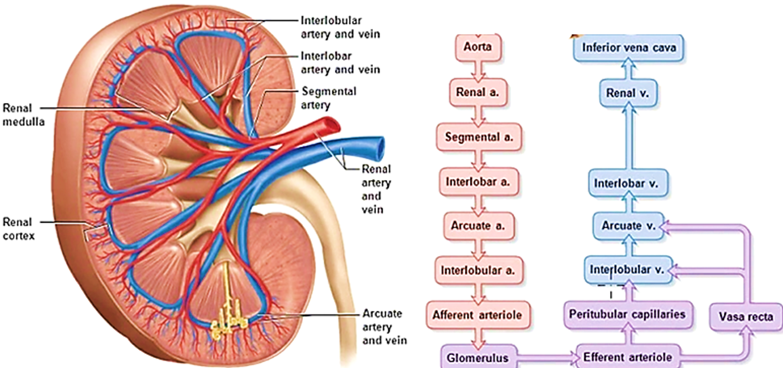
Arterial supply
- Renal arteries arise from abdominal aorta at the level of intervertebral disc between L1 and L2.
- At hilum of the kidney the renal artery divides into 5 segmental branches.
- In the renal sinus, the segmental arteries divide into lobar arteries, which further divide into interlobar arteries.
- Interlobar arteries pass between pyramids in the medulla.
- At the cortico-medullary junction, the each interlobar artery divides into two arcuate arteries (they do not anastomose with adjacent arcuate arteries) .
- From arcuate arteries arise interlobular arteries at right angle and pass through the cortex.
- Many afferent arterioles arise from interlobular artery which feed glomerular capillaries, which in turn end in efferent arteriole.
- Efferent arterioles breakup into peritubular capillary plexus around collecting tubules and ducts.
Venous drainage
- Peritubular plexus drains into interlobular veins, which drain successively into arcuate, interlobular, interlobar, segmental and finally into renal veins.
- Renal veins drain into inferior vena cava.
- Left renal vein receives blood from left suprarenal and left gonadal veins.
Describe the renal vascular segments.
Renal artery usually gives into 5 segmental Based on the distribution of five segmental branches of renal artery, each kidney is divided into five vascular segment.
- Apical/superior
- Upper anterior
- Middle anterior
- Caudal/inferior
- Posterior

Applied Aspects
Perinephric abscess
Pus in a perinephric abscess or blood from a ruptured kidney (perirenal effusions) will first distend the renal fascia and then descend within the fascial compartment downwards into the pelvis. The anterior and posterior layers of renal fascia are fused above, laterally and are connected by a septa medially, therefore the pus cannot pass in these directions.
Floating kidney/ Nephroptosis
Perirenal fascia and fat helps in keeping the kidney in its normal position. In case the perirenal fat is reduced considerably, the kidney may become mobile and descend down towards pelvis.
Renal colic
It is commonly caused by renal stones begins in the loin (flank region) and often radiates to the groin. The kidneys are supplied by T10-L1 spinal segments and the pain to the flank along the subcostal nerve and to the groin along the ilioinguinal nerve.
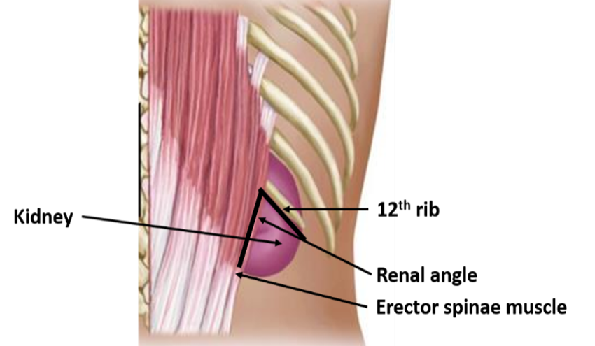
Renal angle
It is the angle between the lower border of the 12th rib and the lateral border of erector spinae muscle. It is the site of tenderness in case of perinephric abscess.

Great, anatomy has been made simpler under one roof.
Thanks a lot.
Great, anatomy has been made quite simpler under this site.
Thanks.
amazing~~~
thank you!!!!!