Advertisements
Describe the location and extent of inguinal canal.
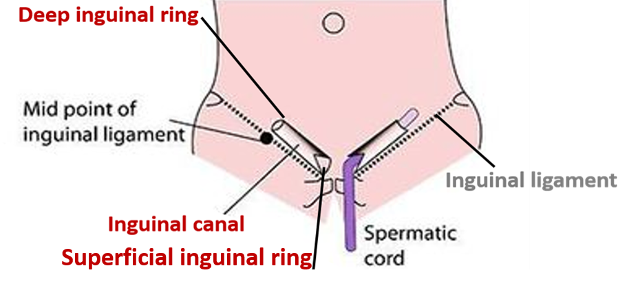
Inguinal canal is an oblique intermuscular passage in the lower part of anterior abdominal wall for the passage of spermatic cord in males and the round ligament of the uterus in the females.

Location:
- It lies ½ inch above and parallel to the medial half of the inguinal ligament.
- In adults, the canal is directed downwards, forwards and medially.
- In newborn, the canal is directed straight forward.
Extent :
- It is about 4cm. long and extends from deep inguinal ring to the superficial inguinal ring.

Describe the boundaries and formation of inguinal canal.
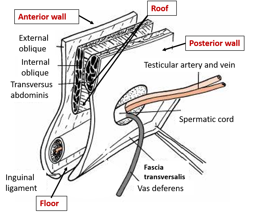
Following are the boundaries of inguinal canal:
- Anterior wall is formed by:
- Skin
- Superficial fascia
- Aponeurosis of external oblique
- In lateral 1/3rd by fleshy fibers of internal oblique
- Posterior wall is formed by:
- Transversalis fascia
- Medial ½ – in front of fascia transversalis by conjoint tendon
- Medial 1/4th – in front of conjoint tendon by reflected part of inguinal ligament.
- Roof is formed by:
- Arched fibers of internal oblique
- Arched fibers of transverses abdominis
- Floor is formed by:
- Grooved upper surface of inguinal ligament
- Medially, upper surface of lacunar ligament

Inlet and outlet:
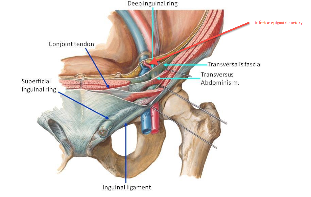
- Inlet is known as deep inguinal ring:
- Is an oval shaped depression (opening) in fascia transversalis.
- Is situated ½ inch above the midinguinal point.
- Is lateral to the inferior epigastric artery.
- Structures passing through are:
- Spermatic cord in males and round ligament of uterus in females.
- Genital branch of genitofemoral nerve
- Outlet is known as superficial inguinal ring:
- Is a triangular gap in the aponeurosis of external oblique , above and lateral to the pubic crest.
- Structures passing through are:
- Spermatic cord in males and round ligament of uterus in females
- Ilioinguinal nerve
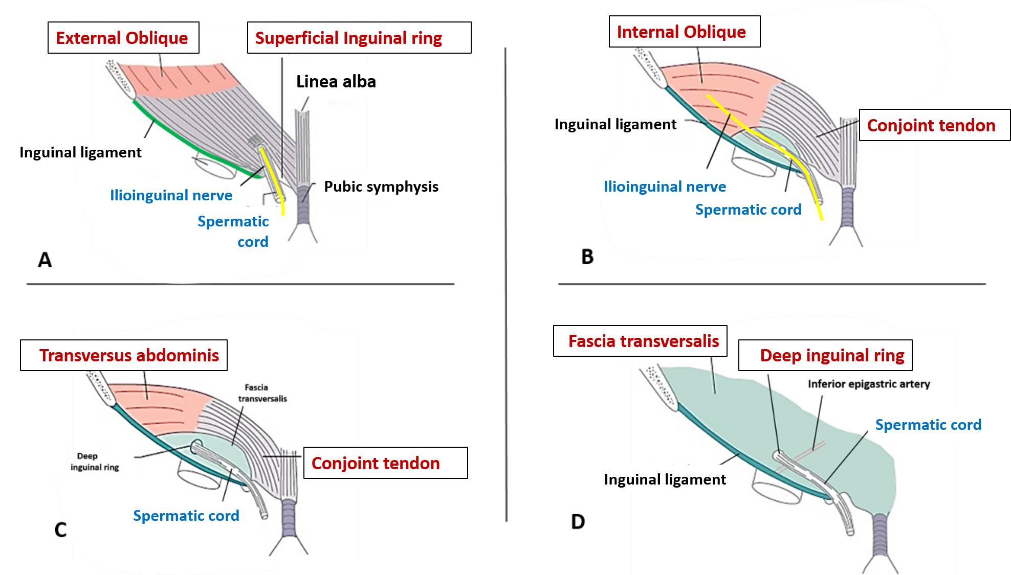
Formation of inguinal canal from outside inwards in males

What are the contents of inguinal canal?
Contents of inguinal canal are:
- In males: spermatic cord and ilioinguinal nerve
- In females: round ligament of uterus and ilioinguinal nerve
Ilioinguinal nerve does not enter the canal through deep inguinal ring but does so by piercing internal oblique muscle and comes out through the superficial inguinal ring.
Describe the protective mechanisms of inguinal canal that prevent inguinal hernia.
Protective mechanisms that prevent inguinal hernia are following:
- Opposite the deep inguinal ring the anterior wall of the canal is strengthened by the fleshy fibers of internal oblique muscle.
- Opposite the superficial inguinal ring the posterior wall is strengthened by conjoint tendon and reflected part of inguinal ligament.
- Obliquity of the canal produces a flap-valve mechanism. During increased intra abdominal pressure the posterior wall comes in contact with anterior wall and the canal is obliterated .
- Contraction of arched fibers of internal oblique and transversus abdominis bring the roof in approximation to the floor like a shutter ( Keith’s shutter mechanism).
- Contraction of external oblique muscles tends to plug the superficial inguinal ring. In males contraction of cremaster muscle pulls up spermatic cord and plugs superficial inguinal ring (Ball –valve mechanism).
- Contraction of external oblique brings the two crura of superficial opening close to each other (Slit valve mechanism).
For Ingunial hernias[Click here]
