Describe the location and boundaries of third ventricle.
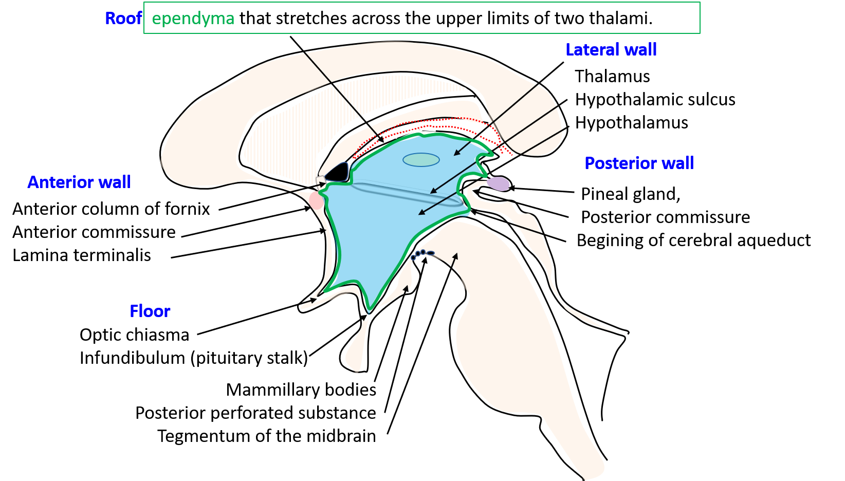
It is a cavity within Diencephalon.It is a midline slit-like cavity situated between the two thalami and the part of hypothalamus.
Boundaries:The third ventricle has
- Anterior wall
- Posterior wall
- Roof
- Floor and
- Two lateral walls.
Anterior wall is formed from above downwards by:
– anterior column of fornix,
– anterior commissure and
– lamina terminalis.

Posterior wall is formed from above downwards by:
– pineal gland,
– posterior commissure and
– begining of cerebral aqueduct.
Roof is formed by: ependyma that stretches across the upper limits of two thalami.
Floor is formed from before backwards by:
– optic chiasma,
– infundibulum (pituitary stalk),
– mammillary bodies,
– posterior perforated substance and
– tegmentum of the midbrain.
Lateral wall is marked by a curved hypothalamic sulcus extending from the interventricular foramen to the cerebral aqueduct.The sulcus divides the
lateral wall into:
i. Upper part formed by the medial surface of the thalamus.
ii. Lower part of the lateral wall is formed by the hypothalamus.
What are the recesses of third ventricle?
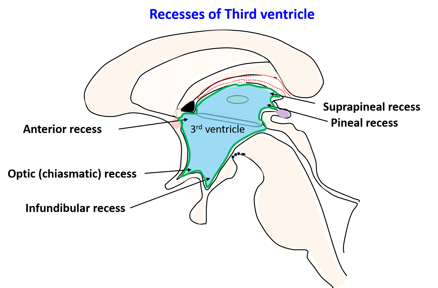
Recesses are pocket-like protrusion of the cavity of third ventricle into the surrounding structures .These are as follows: 1. Infundibular recess: It is a recess that extends downwards into the infundibulum, i.e. the stalk of the pituitary gland.
2. Optic (chiasmatic) recess: It is recess situated at the junction the anterior wall and the floor of the ventricle just above the optic chiasma.\
3. Anterior recess: It is a recess which extends anteriorly in front of interventricular foramen.
4. Suprapineal recess: It is a recess that extends posteriorly above the stalk of pineal gland.
5. Pineal recess: It is recess which extends posteriorly between the superior and inferior laminae of the stalk of the pineal gland.
Describe briefly the tela choroidea and the choroid plexus of third ventricle.

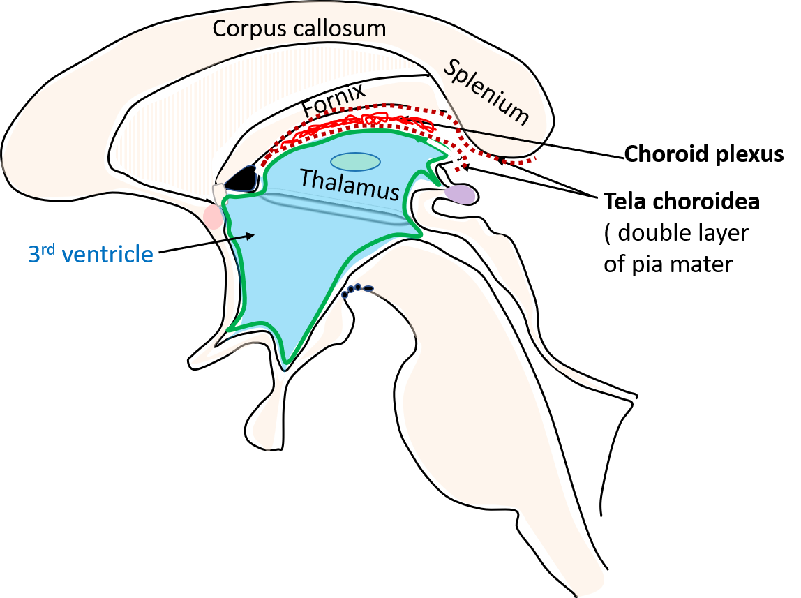
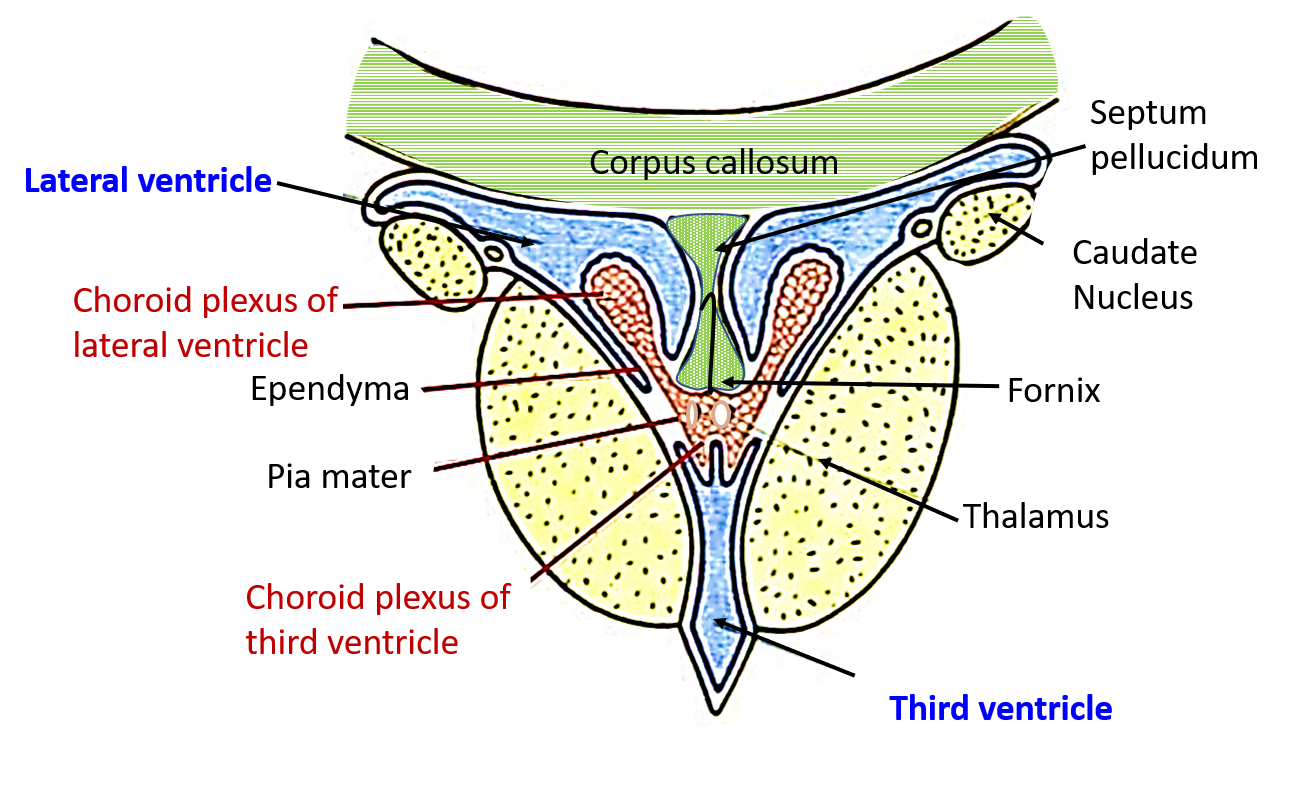
There is a common tela choroidea for third and lateral ventricle. It is a triangular shaped double layer of pia mater which is present in the interval between the splenium of corpus callosum and fornix above and the two thalami below. It extent is anteriorly till the interventricular foramen and posteriorly till the transverse fissure ( gap between the splenium of corpus callosum and roof of 3rd ventricle). The median part lies over the roof of the 3rd ventricle , whereas the lateral margins project through the choroid flissure into the lateral ventricle. The choroid plexus of third ventricle is formed by the capillaries derived from the branches of anterior choroidal arteries which form two antereoposterior longitudinal r vascular fringes between the two layers of pia mater.


THANKYOU FOR THIS!!
THIS SITE IS LIFE SAVER….so glad i found it
I had been struggling to study the 3rd ventricle for a long time, and this has made it super easy to understand. thanks a lot.
Great!!!!
Thanks Ebra.